Page 243 - Veterinary Histology of Domestic Mammals and Birds, 5th Edition
P. 243
Digestive system (apparatus digestorius)   225


       VetBooks.ir


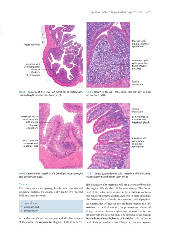
                  10.64  Jejunum at the level of Meckel’s diverticulum.   10.65  Ileum with villi (chicken). Haematoxylin and
                  Haematoxylin and eosin stain (x10).            eosin stain (x40).


                    Intestinal villi in
                     small intestine
                       with simple
                         columnar
                        epithelium


                    Lamina propria
                      mucosae and
                      mucosal folds


                  10.66  Caecum with intestinal villi (chicken). Haematoxylin   10.67  Cloaca (coprodeum) with intestinal villi (chicken).
                  and eosin stain (x25).                         Haematoxylin and eosin stain (x42).


                  Cloaca                                         ible boundary. The intestinal villi are particularly broad in
                  The common excretory passage for the avian digestive and  this region. Distally the villi become shorter. The dorsal
                  urogenital systems, the cloaca, is divided by two mucosal  wall of the subsequent segment, the urodeum, contains
                  folds into three sections:                     the ostia of the paired ureters. Adjacent to these openings,
                                                                 the deferent ducts of male birds open on conical papillae.
                   ·  coprodeum,                                 In females, the left side of the urodeum receives the left
                   ·  urodeum and                                oviduct. In the final section, the proctodeum, the rectal
                   ·  proctodeum.                                lining transitions to a non-glandular mucosa that is con-
                                                                 tinuous with the external skin. The opening of the cloacal
                  In the chicken, the rectum merges with the first segment  bursa (bursa cloacalis, bursa of Fabricius) is in the dorsal
                  of the cloaca, the coprodeum (Figure 10.67) with no vis-  wall of the proctodeum (see Chapter 8, ‘Immune system


       Vet Histology.indb   225                                                                                  16/07/2019   15:01

