
Page 252 - Veterinary Histology of Domestic Mammals and Birds, 5th Edition
P. 252
234 Veterinary Histology of Domestic Mammals and Birds
project into the canaliculi, increasing the capacity for bile
VetBooks.ir excretion. The membrane lining the canaliculi is rich in
membrane-active enzymes (ATP) and resistant to the
effects of bile acids. Intercellular junctions prevent leak-
age of bile into the intercellular space adjacent to the bile
canaliculi.
Bile ducts
The following structures and processes are responsible for
the formation, excretion and transport of bile:
· intracellular production of bile acids in the smooth
ER and Golgi apparatus of the hepatocyte,
· vesicular intracellular transport to the bile canaliculi
(canaliculi biliferi) lined by modified plasmalemma,
· luminal intrahepatic transport of bile through small
bile ductules (ductuli biliferi) lined by cuboidal
epithelium, followed by larger ductules (ductuli inter-
lobulares; one of the components of the portal canal),
· passage of bile into the hepatic duct (ductus
hepaticus),
· luminal extra-hepatic transport of bile acids in the
cystic duct (from the gall bladder) and bile duct and
· delivery of bile into the duodenum (duodenal papilla).
Within liver lobules, bile flows through a network of tubular
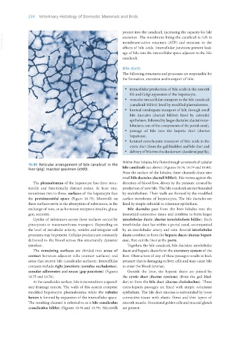
10.80 Reticular arrangement of bile canaliculi in the
liver (pig). Injected specimen (x560). bile canaliculi (see above) (Figures 10.76, 10.79 and 10.80).
Near the surface of the lobules, these channels drain into
small bile ductules (ductuli biliferi). Bile moves against the
The plasmalemma of the hepatocyte has three struc- direction of blood flow, driven by the pressure created by
turally and functionally distinct zones. At least one, production of new bile. The bile canaliculi are not bounded
sometimes two to three, surfaces of the hepatocyte face by endothelium. Their walls are formed by the modified
the perisinusoidal space (Figure 10.79). Microvilli on surface membrane of hepatocytes. The bile ductules are
these surfaces serve in the absorption of substances, in the lined by simple cuboidal to columnar epithelium.
exchange of ions, or as hormone receptors (insulin, gluca- Bile ductules pass from the liver lobules into the
gon, secretin). interstitial connective tissue and combine to form larger
Uptake of substances across these surfaces occurs by interlobular ducts (ductus interlobularis bilifer). Each
pinocytosis or transmembrane transport. Depending on interlobular duct lies within a portal canal, accompanied
the level of metabolic activity, vesicles and irregular cell by an interlobular artery and vein. Several interlobular
processes may be present. Cellular products are constantly ducts combine to form the hepatic ducts (ductus hepati-
delivered to the blood across this structurally dynamic cus), that exit the liver at the porta.
interface. Together, the bile canaliculi, bile ductules, interlobular
The remaining surfaces are divided into areas of ducts and hepatic ducts form the excretory system of the
contact between adjacent cells (contact surfaces) and liver. Obstruction of any of these passages results in back
areas that secrete bile (canalicular surfaces). Intercellular pressure that is damaging to liver cells and may cause bile
contacts include tight junctions (zonulae occludentes), to enter the blood (icterus).
zonulae adherentes and nexus (gap junctions) (Figures Outside the liver, the hepatic ducts are joined by
10.75 and 10.76). the cystic duct (ductus cysticus) (from the gall blad-
At the canalicular surface, bile is excreted into a special- der) to form the bile duct (ductus choledochus). These
ised drainage system. The walls of this system comprise extra-hepatic passages are lined with simple columnar
modified hepatocyte plasmalemma while the tubular epithelium. The bile duct mucosa is surrounded by loose
lumen is formed by expansion of the intercellular space. connective tissue with elastic fibres and thin layers of
The resulting channel is referred to as a bile canaliculus smooth muscle. Occasional goblet cells and mucoid glands
(canaliculus bilifer) (Figures 10.76 and 10.79). Microvilli are present.
Vet Histology.indb 234 16/07/2019 15:02