
Page 253 - Veterinary Histology of Domestic Mammals and Birds, 5th Edition
P. 253
Digestive system (apparatus digestorius) 235
Species variation The tunica mucosa (Figure 10.83) is lined by a simple colum-
VetBooks.ir Birds: The structure of the liver and bile passages of birds reflects the intense absorptive activity of the epithelial cells.
nar epithelium that typically has a distinct brush border. This
is similar to that of mammals. The main distinction is
The prominent intercellular spaces are hyperosmolar (via
the limited amount of interlobular connective tissue, and
thus poor differentiation of hepatic lobules, in birds. The
plates of hepatocytes may be composed of a single layer
(sparrows) or a double layer (chicken) (Figures 10.81 and
10.82). The surface of the avian liver is covered by a tunica
serosa, resting upon a thin stratum fibrosum. The liver
is enclosed by the hepatic peritoneal sac. Double layers of
serous lamellae anchor the liver within the coelomic cavity.
Gall bladder (vesica biliaris, vesica fellea)
The gall bladder stores bile. In addition, bile is concentrated
in the gall bladder by the reabsorption of water. The gall
bladder is absent in the horse. In the empty gall bladder,
the mucosa is thrown into folds (plicae tunicae mucosae).
Invaginations and crypts (cryptae tunicae mucosae) allow
the mucosa to expand without excessive stretching of the
gall bladder wall.
The layers of the gall bladder wall are as follows:
· tunica mucosa: simple columnar epithelium with
microvilli and goblet cells,
· lamina propria mucosae: contains mucoid glands
(particularly in ruminants),
· tunica muscularis: musculo-elastic tissue, smooth
muscle arranged in spiral lattices,
· tunica adventitia: where gall bladder adjoins the
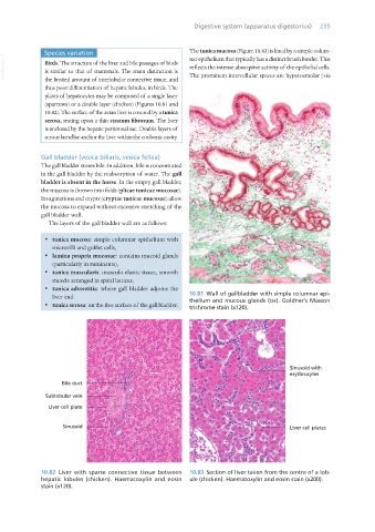
liver and 10.81 Wall of gallbladder with simple columnar epi-
thelium and mucous glands (ox). Goldner’s Masson
· tunica serosa: on the free surface of the gall bladder. trichrome stain (x120).
10.82 Liver with sparse connective tissue between 10.83 Section of liver taken from the centre of a lob-
hepatic lobules (chicken). Haematoxylin and eosin ule (chicken). Haematoxylin and eosin stain (x200).
stain (x120).
Vet Histology.indb 235 16/07/2019 15:02