Page 258 - Veterinary Histology of Domestic Mammals and Birds, 5th Edition
P. 258
240  Veterinary Histology of Domestic Mammals and Birds


       VetBooks.ir


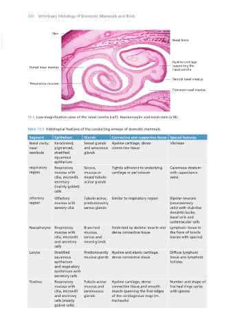
                   11.1  Low-magnification view of the nasal concha (calf). Haematoxylin and eosin stain (x10).


                   Table 11.1  Histological features of the conducting airways of domestic mammals.

                    Segment      Epithelium     Glands        Connective and supportive tissue Special features
                    Nasal cavity:  Keratinised,   Sweat glands   Hyaline cartilage, dense   Vibrissae
                    nasal        pigmented,     and sebaceous  connective tissue
                    vestibule    stratified     glands
                                 squamous
                                 epithelium
                    respiratory   Respiratory   Serous,       Tightly adherent to underlying   Cavernous stratum
                    region       mucosa with    mucous or     cartilage or periosteum       with capacitance
                                 cilia, microvilli,   mixed tubulo-                         veins
                                 secretory      acinar glands
                                 (mainly goblet)
                                 cells
                    olfactory    Olfactory      Tubulo-acinar,   Similar to respiratory region  Bipolar neurons
                    region       mucosa with    predominantly                               (neurosensory
                                 sensory cilia  serous glands                               cells) with club-like
                                                                                            dendritic bulbs,
                                                                                            basal cells and
                                                                                            sustentacular cells
                    Nasopharynx  Respiratory    Branched      Underlaid by skeletal muscle and  Lymphatic tissue in
                                 mucosa with    mucous,       dense connective tissue       the form of tonsils
                                 cilia, microvilli   serous and                             (varies with species)
                                 and secretory   mixed glands
                                 cells
                    Larynx       Stratified     Predominantly  Hyaline and elastic cartilage,   Diffuse lymphoid
                                 squamous       mucous glands dense connective tissue       tissue and lymphoid
                                 epithelium                                                 follicles
                                 and respiratory
                                 epithelium with
                                 secretory cells
                    Trachea      Respiratory    Tubulo-acinar   Hyaline cartilage, dense    Number and shape of
                                 mucosa with    mucous and    connective tissue and smooth   tracheal rings varies
                                 cilia, microvilli   seromucous   muscle spanning the free edges   with species
                                 and secretory   glands       of the cartilaginous rings (m.
                                 cells (mainly                trachealis)
                                 goblet cells)


       Vet Histology.indb   240                                                                                  16/07/2019   15:02

