
Page 262 - Veterinary Histology of Domestic Mammals and Birds, 5th Edition
P. 262
244 Veterinary Histology of Domestic Mammals and Birds
The structure of the neurosensory olfactory mucosa
region of chickens and water birds comprises a small
VetBooks.ir circumscribed area on the caudal nasal concha and the is similar to that of the olfactory region of the nasal cavity.
Poorly myelinated fibres of the terminal nerve (n. terminalis)
caudal portion of the nasal septum. Both the olfactory
traverse the connective tissue of the lamina propria. These
region and the olfactory bulb of birds are relatively
small and limited in function. In comparative terms, sensory fibres pass to the area cribrosa and then to the
the olfactory system is more developed in carnivores olfactory bulb. The exterior of the vomeronasal organ is
(including fish eaters) than in granivorous species. surrounded by a cuff of hyaline cartilage (vomeronasal
cartilage).
Paranasal sinuses (sinus paranasales) Pharynx
The paranasal sinuses are lined with a low respiratory epithe- The dorsal compartment of the pharynx, the nasophar-
lium. Glands are rarely present. The lamina propria is closely ynx (pars nasalis pharyngis) is lined with respiratory
associated with the periosteum. In carnivores this layer con- mucosa. The lamina propria and tela submucosa contain
tains tubulo-acinar serous glands (in the maxillary sinus). abundant lymphoid nodules. Nodular aggregates form the
Their secretions pass from the sinus into the nasal cavity. pharyngeal tonsils. The lymphoid tissue is immediately
surrounded by tubulo-acinar, predominantly mixed glands.
Vomeronasal organ (organum vomeronasale) The tunica muscularis is composed of skeletal muscle. On
The vomeronasal organ aids in identification of scents and the outer surface of its muscular wall, the nasopharynx
odours. It bears receptors for specific pheromones and is is lined by a dense tunica adventitia. At the level of the
believed to play a role in detecting oestrus, particularly in soft palate (structure described in Chapter 10, ‘Digestive
lower order vertebrates. In domestic mammals, the vom- system’), the nasopharynx is continued by the orophar-
eronasal organ is relatively poorly developed. ynx, in which the respiratory and digestive tracts intersect.
The vomeronasal organ is a paired tubular structure, In this portion of the pharynx, the epithelium is stratified
lying parallel to the base of the nasal septum (Figure 11.5). squamous. Mixed glands and lymphoid tissue are located
Its rostral opening empties into the nasal and oral cavities in the submucosa. The submucosa is permeated by a dense
via the incisive duct. In the horse, the connection to the meshwork of elastic fibres that strengthens the pharyn-
oral cavity is lacking. The caudal end of the vomeronasal geal wall. Lying external to the submucosa are the striated
organ is blind. bundles of the pharyngeal muscles, followed by a layer of
The lumen of the vomeronasal organ is lined with res- fibro-elastic tissue.
piratory mucosa on its lateral aspect and with olfactory
mucosa on its medial internal surface (Figure 11.6).
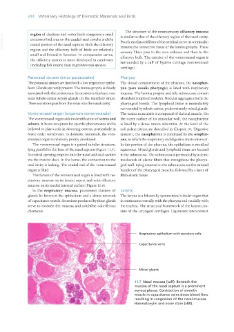
In the respiratory mucosa, prominent clusters of Larynx
glands lie between the epithelium and a dense network The larynx is a bilaterally symmetrical tubular organ that
of capacitance vessels. Secretions produced by these glands is continuous rostrally with the pharynx and caudally with
serve to moisten the mucosa and solubilise odoriferous the trachea. The structural framework of the larynx con-
chemicals. sists of the laryngeal cartilages. Ligaments interconnect
11.7 Nasal mucosa (calf). Beneath the
mucosa of the nasal septum is a prominent
venous plexus. Contraction of smooth
muscle in capacitance veins slows blood flow
resulting in congestion of the nasal mucosa.
Haematoxylin and eosin stain (x80).
Vet Histology.indb 244 16/07/2019 15:02