
Page 260 - Veterinary Histology of Domestic Mammals and Birds, 5th Edition
P. 260
242 Veterinary Histology of Domestic Mammals and Birds
VetBooks.ir
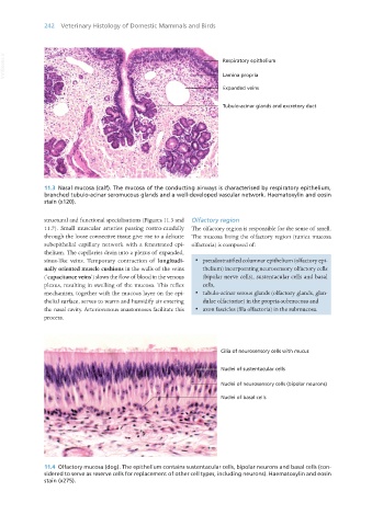
11.3 Nasal mucosa (calf). The mucosa of the conducting airways is characterised by respiratory epithelium,
branched tubulo-acinar seromucous glands and a well-developed vascular network. Haematoxylin and eosin
stain (x120).
structural and functional specialisations (Figures 11.3 and Olfactory region
11.7). Small muscular arteries passing rostro-caudally The olfactory region is responsible for the sense of smell.
through the loose connective tissue give rise to a delicate The mucosa lining the olfactory region (tunica mucosa
subepithelial capillary network with a fenestrated epi- olfactoria) is composed of:
thelium. The capillaries drain into a plexus of expanded,
sinus-like veins. Temporary contraction of longitudi- · pseudostratified columnar epithelium (olfactory epi-
nally oriented muscle cushions in the walls of the veins thelium) incorporating neurosensory olfactory cells
(‘capacitance veins’) slows the flow of blood in the venous (bipolar nerve cells), sustentacular cells and basal
plexus, resulting in swelling of the mucosa. This reflex cells,
mechanism, together with the mucous layer on the epi- · tubulo-acinar serous glands (olfactory glands, glan-
thelial surface, serves to warm and humidify air entering dulae olfactoriae) in the propria-submucosa and
the nasal cavity. Arteriovenous anastomoses facilitate this · axon fascicles (fila olfactoria) in the submucosa.
process.
11.4 Olfactory mucosa (dog). The epithelium contains sustentacular cells, bipolar neurons and basal cells (con-
sidered to serve as reserve cells for replacement of other cell types, including neurons). Haematoxylin and eosin
stain (x275).
Vet Histology.indb 242 16/07/2019 15:02