
Page 44 - Veterinary Histology of Domestic Mammals and Birds, 5th Edition
P. 44
26 Veterinary Histology of Domestic Mammals and Birds
HETEROCHROMATIN (HETEROCHROMATINUM) particularly dense and homogeneous, and are strongly
VetBooks.ir In heterochromatin, large portions of chromatin refractive. They can be subdivided into three regions:
are strongly coiled and folded (Figures 1.6 and 1.33).
Histologically, heterochromatic regions of the nucleus
· granular component (pars granulosa): consists pri-
appear irregularly granular, are frequently darkly staining marily of pre-ribosomal RNA particles (10–15 nm);
(basophilic) and can be selectively identified using various peripherally located,
histological techniques. A high proportion of heterochro- · dense fibrillar component (pars fibrosa): contains
matin signifies a relatively low level of cellular metabolic RNA transcripts, usually forms central clumps and
activity. Heterochromatin is predominantly located · fibrillar centres: regions containing DNA compo-
peripherally but can also be widely distributed through- nents (intra- and perinucleolar chromatin) involved
out the nucleus. in RNA synthesis.
Heterochromatin is not under hormonal control, is sen-
sitive to breakage and exhibits a high rate of mutation. In The fibrillar and granular components form a sponge-like
contrast to euchromatin, it is highly resistant to the effects network (nucleolonema). Intra- and perinucleolar chro-
of radiation (e.g. X- and UV radiation). matin is present in the spaces between the anastomosing
Sex chromatin represents a specialised form of chro- strands of the nucleolonema. Some cells (e.g. oocytes)
matin found in female cells. This component of the have a ring-shaped nucleolus in which there is a distinct
X-chromosome remains condensed during interphase and separation of the two RNA components.
can be identified histologically as a typically peripherally The nucleolus is a centre of RNA synthesis. Ribosomes
located region of dense chromatin or, in neutrophils, as a are produced here and, once assembled, are released
drumstick-shaped nuclear appendage. into the cytoplasm. The size of the nucleolus reflects the
While the amount of euchromatin and heterochro- amount of stored RNA and is a morphological indica-
matin varies from cell to cell, the relative proportion of tor of cellular protein biosynthesis. As synthetic activity
each type of chromatin is consistent within particular cell increases, the granular components of the nucleolus
populations and can thus be used as a diagnostic aid. Cells expand. The presence of several nucleoli in one nucleus
with a ‘light’ (euchromatic) nucleus are usually metaboli- reflects a high level of protein metabolism (e.g. in nerve
cally active, while those with a ‘dark’ (heterochromatic) cells, pancreatic cells, embryonic cells and tumour cells).
nucleus are relatively quiescent.
Cell growth and division
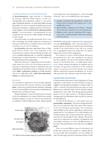
Nucleolus Cellular growth and division are defining features of living
The nucleolus is a spherical to ovoid, strongly basophilic organisms. As the lifespan of each cell is finite, it must be
nuclear inclusion that is present exclusively during inter- equipped with the genetic machinery to replicate itself. In
phase. It arises after mitosis from the nucleolar organising order to maintain vital physiological processes, an equi-
centre of various different chromosomes and disintegrates librium exists within the body between cell death and the
shortly before cell division begins. production of new cells. Only during physical growth,
Nucleoli (diameter 3 μm) are found as free structures phases of cell or organ renewal, or during physiological
within the nucleoplasm or in contact with the inner nuclear derangements does this balance shift towards a net increase
membrane (Figures 1.6 and 1.36). They are not enclosed by in cell production.
a membrane. With the light microscope, nucleoli appear The bulk of the lifespan of a cell is taken up by the
growth phase (interphase). The period during which divi-
sion occurs accounts for only a few hours of the existence
of the cell. During the growth phase, the cell increases
(often doubles) in size, and its organelles are replicated, in
preparation for the production of a viable daughter cell.
During this phase, the cell also performs the various func-
tions that are characteristic for that cell type. The growth
phase thus incorporates the synthesis of endogenous and
extracellular substances, as well as cell differentiation. At
the end of the growth phase, most cells enter the mitotic
phase.
There are several different types of cell division. By
far the most common is the division of a mother cell into
two genetically identical daughter cells that exhibit the
1.36 Electron microscope image of a nucleolus (x28,000). same morphological and functional characteristics. In this
Vet Histology.indb 26 16/07/2019 14:53