
Page 39 - Veterinary Histology of Domestic Mammals and Birds, 5th Edition
P. 39
The cell (cellula) 21
superficial cells subjected to large mechanical forces (sur- Exogenous and endogenous cellular
VetBooks.ir myofibril bundles (at the Z line) (Table 1.1). Exogenous inclusions are taken up from the exterior of
face epithelia), in axons and at junctions between adjacent inclusions
Intermediate filament-associated proteins play an the cell (e.g. by phagocytosis). Endogenous inclusions
important role in the cytoskeleton. Some (e.g. plectins) are those formed within the cell as by-products of cellu-
bind to actin filaments and microtubules while others lar metabolism. Together, these inclusions are sometimes
(e.g. desmoplakins or plakoglobulins) form attachment termed paraplasm. They are highly heterogeneous
plaques for the cytoskeleton, which are constituents of (Figures 1.31 and 1.32) and are subdivided into:
desmosomes and hemidesmosomes.
In contrast to actin filaments and microtubules, inter- · storage and reserve materials (e.g. glycogen, fats,
mediate filaments do not disassemble and reform. Instead, proteins) and
they constitute substantial, permanent structural ele- · pigments:
ments of the cell. They contribute to the integrity of − endogenous (e.g. ferritin, haemosiderin, mela-
cell-to-cell junctions and to connections between the nin) and
cell and the extracellular matrix. The composition of the − exogenous (e.g. dust, heavy metals).
polypeptide chain of intermediate filaments exhibits con-
siderable cell specificity. Several classes are recognised Characteristic inclusions include intracellular fat droplets,
(Table 1.2), including: glycogen (Figure 1.31), protein crystals and pigments.
Pigments (see below) represent a special form of inclusion.
· Class I and II: keratins,
· Class III: vimentin and vimentin-like filaments,
· Class IV: neurofilaments and
· Class V: lamins.
Keratins (cytokeratins, tonofilaments)
This group includes more than 50 different cell- and tis-
sue-specific isoforms and subtypes. Keratins are found
particularly in cells of epithelial origin. Their component
keratin proteins form an intracellular network, organ-
ised along mechanical lines, that inserts on desmosomes
at the plasmalemma. Through these cellular junctions,
the filaments extend functionally into neighbouring cells
and serve to increase the strength of the epithelium as a
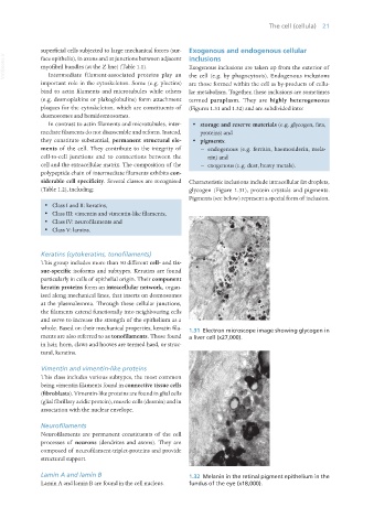
whole. Based on their mechanical properties, keratin fila- 1.31 Electron microscope image showing glycogen in
ments are also referred to as tonofilaments. Those found a liver cell (x27,000).
in hair, horn, claws and hooves are termed hard, or struc-
tural, keratins.
Vimentin and vimentin-like proteins
This class includes various subtypes, the most common
being vimentin filaments found in connective tissue cells
(fibroblasts). Vimentin-like proteins are found in glial cells
(glial fibrillary acidic protein), muscle cells (desmin) and in
association with the nuclear envelope.
Neurofilaments
Neurofilaments are permanent constituents of the cell
processes of neurons (dendrites and axons). They are
composed of neurofilament-triplet-proteins and provide
structural support.
Lamin A and lamin B 1.32 Melanin in the retinal pigment epithelium in the
Lamin A and lamin B are found in the cell nucleus. fundus of the eye (x18,000).
Vet Histology.indb 21 16/07/2019 14:53