
Page 37 - Veterinary Histology of Domestic Mammals and Birds, 5th Edition
P. 37
The cell (cellula) 19
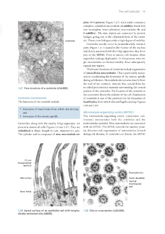
plets (9×3 pattern) (Figure 1.27). Each triplet contains a
VetBooks.ir complete, cylindrical microtubule (A-subfibre) fused with
two incomplete hemi-cylindrical microtubules (B- and
C-subfibre). The nine triplets are connected by protein
bridges, giving rise to the cylindrical form of the centri-
ole. These cross-linkages confer a high degree of stability.
Centrioles usually occur in perpendicularly oriented
pairs (Figure 1.6). Located in the vicinity of the nucleus
and closely associated with the Golgi apparatus, they form
part of the MTOC. Prior to mitotic cell division, these
organelles undergo duplication. In this process, nine sin-
gle microtubules are formed initially; these subsequently
expand into triplets.
The known functions of centrioles include organisation
of intracellular microtubules. This is particularly impor-
tant in coordinating the formation of the mitotic spindle
during cell division. Microtubules do not arise directly from
the wall of the centriole. Instead, they extend from the
1.27 Fine structure of a centriole (x34,000). so-called pericentriolar material surrounding the central
portion of the centrioles. The location of the centriole in
the cytocentre directs the polarity of the cell. Duplication
Centriole (centriolum) of centrioles is one of the pathways for the formation of
The functions of the centriole include: basal bodies, from which cilia and flagella develop (Figures
1.28 and 1.29).
· formation of basal bodies from which cilia develop
and Microtubule-organising centre (MTOC)
· formation of the mitotic spindle. The microtubule-organising centre (cytocentre, cen-
trosome) incorporates both the centrioles and the
Centrioles, along with the nearby Golgi apparatus, are pericentriolar material. Most microtubules are associated
present in almost all cells (Figures 1.6 and 1.27). They are with the MTOC. The MTOC controls the number, polar-
cylindrical in shape (length 0.3 μm, diameter 0.1 μm). ity, direction and organisation of microtubules formed
The cylinder wall is composed of nine microtubule tri- during cell division. If centrioles are absent, the MTOC
1.28 Apical surface of an epithelial cell with longitu- 1.29 Cilia in cross-section (x20,000).
dinally sectioned cilia (x8000).
Vet Histology.indb 19 16/07/2019 14:53