
Page 33 - Veterinary Histology of Domestic Mammals and Birds, 5th Edition
P. 33
The cell (cellula) 15
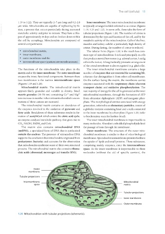
1.19 to 1.22). They are typically 2–7 μm long and 0.2–2.0 Inner membrane: The inner mitochondrial membrane
VetBooks.ir μm wide. Mitochondria are capable of replicating by fis- is typically arranged in folds referred to as cristae (Figures
sion, a process that occurs particularly during increased 1–19, 1.21 and 1.22). In some cells, these take the form of
tubular projections (Figure 1.20). The number of cristae is
metabolic activity and prior to mitosis. They have a lifes-
pan of approximately 20 days and are broken down within determined by the type and function of the cell, and by the
the cell by autophagy. Mitochondria are composed of metabolic activity of the mitochondrion. Cells of the liver,
several compartments: muscles and kidney exhibit a particularly high density of
cristae. During fasting, the number of cristae is reduced.
· mitochondrial matrix, The tubular form (Figure 1.20) is the much less com-
· inner membrane, mon type of mitochondrion. It is found principally in cells
· outer membrane and the that produce steroid hormones (e.g. adrenal cortex, Leydig
· intermembrane space (spatium intermembranosum). cells in the testes). A longitudinally prismatic arrangement
of the cristal membrane is also recognised (e.g. glial cells).
The functions of the mitochondria take place in the The inner mitochondrial membrane contains a large
matrix and at the inner membrane. The outer membrane number of enzymes that are essential for sustaining life,
encases the inner, functional components. Between these a feature that distinguishes it from other cell membranes.
two membranes is the narrow intermembrane space On the surface facing the matrix, the membrane carries
(Figures 1.19 and 1.20). enzymes associated with the respiratory chain (electron
Mitochondrial matrix: The mitochondrial matrix transport chain) and oxidative phosphorylation. The
appears finely granular and variable in density. Small vast majority of energy for the cell is generated at the inner
2+
2+
matrix granules (30–50 nm) containing Ca and Mg mitochondrial membrane, through the formation of ATP
ions increase in number when intramitochondrial concen- from adenosine diphosphate (ADP) and inorganic phos-
trations of these cations are increased. phate. The morphological entities associated with energy
The mitochondrial matrix contains an abundance of generation, referred to as elementary particles, consist of
the enzymes involved in the oxidation of pyruvate and a globular enzyme-containing head and a stalk connected
fatty acids. Breakdown of these substrates results in for- to the inner membrane by a foot-plate (Figure 1.23) (refer
mation of acetyl-CoA which enters the citric acid cycle, to biochemistry texts for further detail).
an enzyme-catalysed metabolic pathway that gives rise to The inner mitochondrial membrane is impermeable to
CO , NADH, FADH and CoA. many molecules. Abundant embedded phospholipids limit
2 2
The matrix also contains mitochondrial DNA the passage of ions through the membrane.
(mtDNA), a specialised form of DNA that is synthesised Outer membrane: The structure of the outer mito-
outside the nucleus. The presence of extranuclear DNA chondrial membrane is similar to that of other biological
supports the conclusion that mitochondria originated from membranes. Specialised transmembrane proteins facilitate
prokaryotes (bacteria) and accounts for the observation the uptake of lipids and small proteins. These substances,
that mitochondria synthesise some of their own structural comprising mainly enzymes, enter the intermembrane
proteins. The mitochondrial matrix also contains ribonu- space. As the inner membrane is impermeable to these
cleic acids (ribosomal, messenger and transfer RNA). molecules (without the aid of specific carriers), the
1.20 Mitochondrion with tubular projections (schematic).
Vet Histology.indb 15 16/07/2019 14:53