
Page 36 - Veterinary Histology of Domestic Mammals and Birds, 5th Edition
P. 36
18 Veterinary Histology of Domestic Mammals and Birds
subunits: α- and β-tubulin (each molecular weight 50,000
VetBooks.ir Da).
The tubulin dimers (formed from free cytoplasmic
α- and β-tubulin molecules) polymerise end-to-end, the
α-subunit of one molecule binding to the β-component of
another. The resulting chains are termed protofilaments
(Figure 1.30). Thirteen protofilaments combine to form
the wall of the microtubule.
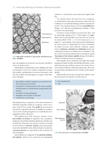
Formation of microtubules is promoted by Mg and
2+
2+
by calmodulin binding of Ca . Microtubules are highly
labile (dynamically unstable) structures that can quickly
be disassembled (Table 1.1). At low temperatures, or in
high Ca environments, they spontaneously depolymer-
2+
ise and can then be reassembled into new microtubules
in another location, under different conditions. Agents
such as colchicine, colcemid and vinblastine prevent the
ordered polymerisation of tubulin into microtubules. They
are used in research and therapeutics as antimitotic agents
1.25 Microvilli covered in glycocalyx (transverse sec- (formation of the mitotic spindle during cell division is
tion, x40,000). inhibited, blocking mitosis in metaphase).
Microtubules can be identified with light microscopy
new microtubules are formed in one location; elsewhere, using special stains (labelled anti-tubulin antibodies), polar-
others are broken down. isation microscopy and phase contrast microscopy. Due to
Microtubules are inherently flexible. Binding with other their limited resolution under light microscopy, they may
microtubules or with other cellular components increases erroneously be described as fibres or fibrils (e.g. the mitotic
their rigidity, contributing to the structural integrity of the spindle).
cell. Microtubules also participate in a range of other func- Microtubules can become arranged into ordered, com-
tions including: plex structures, forming the structural basis of:
· intracellular vesicular transport (e.g. secretory vesi- · the centriole and
cles, endosomes, lysosomes), · cilia.
· attachment of chromosomes to the mitotic spindle
and movement of chromosomes during mitosis and
meiosis,
· movement of cilia and flagella,
· elongation and motility of cells and
· maintenance of cell shape.
Microtubules play an important role in the movement of
individual organelles within the cytoplasm, and in move-
ment of the cell as a whole. They guide the movement of
intracellular organelles and serve as ‘tracks’ for transport
vesicles and vacuoles passing between metabolically active
organelles and the cell surface.
Microtubule-associated transport proteins direct
intracellular movement of organelles and cytoplasmic
inclusions towards their intended destination. Entities des-
tined for the cell surface (e.g. secretory vesicles) are bound
to microtubules by kinesin. Dynein carries intracellular
structures towards the centre of the cell and to the nucleus.
Axonemal dyneins are responsible for the movement of
cilia and flagella. 1.26 Fine structure of microtubules (x10,000).
Microtubules are composed of the globular polypep-
tide tubulin, which in turn consists of two polypeptide
Vet Histology.indb 18 16/07/2019 14:53