
Page 42 - Veterinary Histology of Domestic Mammals and Birds, 5th Edition
P. 42
24 Veterinary Histology of Domestic Mammals and Birds
VetBooks.ir
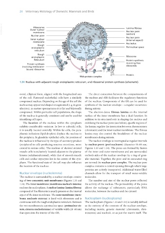
1.34 Nucleus with adjacent rough endoplasmic reticulum, and ribosomal protein synthesis (schematic).
ovoid, elliptical form, aligned with the longitudinal axis The direct connection between the compartments of
of the cell. Flattened endothelial cells have a similarly the nucleus and rER facilitates the regulatory functions
compressed nucleus. Depending on the age of the cell the of the nucleus. Components of the ER can be used for
nucleus may appear rod-shaped or segmented (e.g. in gran- synthesis of the nuclear envelope – a regular occurrence
ulocytes); in mature spermatozoa it is flat and bilaterally during mitosis.
compressed. Within particular cell populations, the shape The electron-dense fibrous lamina on the internal
of the nucleus is generally consistent and can be used for surface of the inner membrane has a dual function. In
identifying cell types. addition to its structural role in shaping the nucleus and
The location of the nucleus within the cytoplasm stabilising the nuclear pores (see below), specific regions of
exhibits considerable variation. In free or cuboidal cells, the lamina regulate the interaction between chromosomes
it is usually located centrally. Within fat cells, the para- (chromatin) and the inner nuclear membrane. The fibrous
plasmic inclusions (lipid droplets) displace the nucleus to lamina may also control the breakdown of the nuclear
the periphery. In glandular epithelial cells, the position of membranes during mitosis.
the nucleus is influenced by the type of secretory product The nuclear envelope is interrupted at regular intervals
(peripheral in cells producing mucous secretions, more by nuclear pores (pori nucleares) (diameter 50–80 nm;
central in serous cells). The nucleus of skeletal striated Figures 1.33 and 1.34). The pores are formed by fusion
muscle cells is exclusively located adjacent to the plasma- of the inner and outer membranes and are surrounded
lemma (subplasmalemmal), while that of smooth muscle on both sides of the nuclear envelope by a ring of gran-
cells and cardiac myocytes lies in the centre of the cyto- ular material. Together, the pore and its associated ring
plasm. The functional state of the cell may also influence are termed the nuclear pore complex. The nuclear pore
the location of the nucleus. complex contains a central opening through which large
proteins are actively transported. Additional water-filled
Nuclear envelope (nucleolemma) channels allow for the transport of small water-soluble
The nucleus is surrounded by a nuclear envelope consist- molecules.
ing of two concentric unit membranes (Figures 1.33 and The number and size of the nuclear pores reflected
1.34). The inner membrane (membrana nuclearis interna) the activity of the nucleus. The permeability of the pores
encloses the nucleoplasm. A nuclear lamina (lamina fibrosa) allows the exchange of substances, particularly RNA
composed of fine filaments is usually present on the internal molecules, between the nucleus and the cytosol.
aspect of the inner membrane. The outer membrane (mem-
brana nuclearis externa) is lined with ribosomes and is Nucleoplasm (nucleoplasma)
continuous with the rough endoplasmic reticulum. Between The nucleoplasm (Figures 1.33 and 1.34) is variably defined
the two membranes is a perinuclear space (perinuclear cis- as the entirety of the contents of the nuclear envelope,
tern, cisterna nucleolemmae) of variable width (20–60 nm) including matrix, genetic material (chromatin, chro-
that opens into the interior of the rER. mosomes) and nucleoli, or as just the matrix itself. The
Vet Histology.indb 24 16/07/2019 14:53