
Page 64 - Veterinary Histology of Domestic Mammals and Birds, 5th Edition
P. 64
46 Veterinary Histology of Domestic Mammals and Birds
classification of this epithelium as non-keratinised, the The basal stratum (stratum basale) is composed of
VetBooks.ir superficial cells may contain some keratin. However, unlike basophilic, cuboidal to columnar cells that are firmly
the outermost cells of keratinised epithelium, they retain connected to the basal lamina by hemidesmosomes. The
their nucleus. As this type of epithelium is prone to dehy-
epithelium is thus securely anchored to the underlying
dration, it is found in locations that experience relatively connective tissue. Varying numbers of melanin granules
weak mechanical forces and are kept moist by glandular may be present within the cytoplasm of basal cells. These
secretions. Depending on species, diet and other factors, cells (melanocytes) are responsible for pigmentation of the
these may include the oral cavity, pharynx, oesophagus, epithelium. The stratum basale also contains specialised
anal canal and vagina (Figures 2.13 to 2.15). cells that have immunological and/or phagocytic activity
(Langerhans cells) as well as tactile cells (Merkel cells).
keRatinised stRatified squamous ePithelium The stratum spinosum (prickle cell or spinous layer)
(ePithelium stRatificatum squamosum coRnificatum) is composed primarily of cuboidal polygonal cells that
Keratinised stratified squamous epithelium forms the are tightly interconnected by numerous desmosomes
outer layer of the skin, the epidermis (see also Chapter (maculae adherentes), located at the end of cytoplasmic
15, ‘Common integument’). It consists of up to five dis- extensions. Under light microscopy this gives the cells a
tinct strata that become keratinised towards the surface spiny appearance, from which this layer derives its name.
and vary in their thickness depending upon the loca- This effect is often enhanced by the presence of large
tion of the epithelium within the body (Figures 2.16 and intercellular spaces and a high concentration of intracyto-
2.17). plasmic tonofilaments. The latter are arranged in a lattice
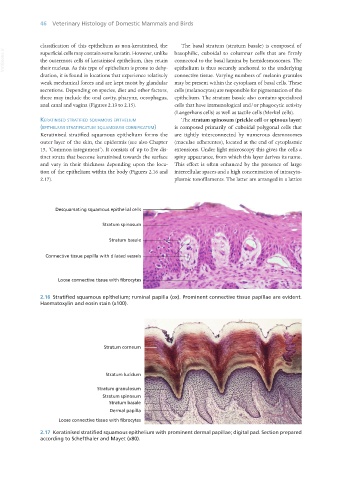
Desquamating squamous epithelial cells
Stratum spinosum
Stratum basale
Connective tissue papilla with dilated vessels
Loose connective tissue with fibrocytes
2.16 Stratified squamous epithelium; ruminal papilla (ox). Prominent connective tissue papillae are evident.
Haematoxylin and eosin stain (x100).
2.17 Keratinised stratified squamous epithelium with prominent dermal papillae; digital pad. Section prepared
according to Schefthaler and Mayet (x80).
Vet Histology.indb 46 16/07/2019 14:54