Page 256 - Zoo Animal Learning and Training
P. 256
240    Tasks for the Veterinary Assistant


                                                             the surface of the agar. Each disk is coded with the anti-
                                                             biotic it contains. Compare the code on the disk with the
                                                             code on the tube to determine the name of the antibi-
                                                             otic it contains. The disks are pressed into the agar with
                                                             a flamed loop or sterile swab. The dish is inverted,
                                                             labeled, and incubated.
                                                               The antibiotic seeps out into the agar surrounding
                                                             the disk. After incubation, the plate is held to a bright
                                                             light and zones of inhibition of bacterial growth are
                                                             noted and measured around each disk. If the bacteria
                                                             are sensitive to the antibiotic, they will not grow up to the
                                                             disk. The diameter sizes of the zones are compared with
                                                             the manufacturer’s standardized  chart to determine
                                                             which antibiotic is the best. Some bacteria will be resis-
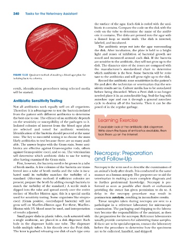
          FIGURE 12.22  Quadrant method of steaking a blood agar plate for   tant to the antibiotics and will grow right up to the disk.
          isolating bacteria colonies.
                                                               Record the antibiotic zone sensitivities in the patient’s
                                                             file and alert the technician or veterinarian that the sen-
          result, identification procedures using selected media   sitivity results are in. Culture media has to be autoclaved
          will be started.                                   before being discarded. When a Petri dish is no longer
                                                             needed place it in an autoclavable bag. Seal the bag with
          Antibiotic Sensitivity Testing                     autoclave tape and run it through a general autoclave
                                                             cycle to destroy all of the bacteria. Then it can be dis-
          Not all antibiotics work equally well on all organisms.   posed of in the regular garbage.
          Therefore it is advantageous to test the bacteria isolated
          from the patient with different antibiotics to determine
          the best one to use. The efficacy of an antibiotic depends
          on the sensitivity or susceptibility of the pathogen to it.   Learning Exercise
          Isolated colonies of interest from the blood agar plate   If available look at the antibiotic disk dispenser.
          are selected and tested for antibiotic sensitivity.   Write down the types of antibiotics available, then
          Identification of the bacteria should proceed at the same   look them up on the internet.
          time. The key to successful testing is to choose the most
          likely antibiotics to test because there are so many avail-
          able. The answer begins with the Gram stain. Some anti-
          biotics are effective against Gram‐negative rods, others
          against Gram‐positive cocci, and so on. The veterinarian   Necropsy: Preparation
          will determine which antibiotic disks to use for testing
          after having examined the Gram stain.              and Follow‐Up
            First, however, the bacteria need to be grown in a tube
          of broth media. A few colonies of the bacteria are trans-  Necropsy is the term used to describe the examination of
          ferred into a tube of broth media and the tube is incu-  an animal’s body after death. It is conducted in the same
          bated until its turbidity matches the turbidity of a   manner as a human autopsy. The purposes are to aid the
          standard. (Alternate method: if sufficient colonies are   veterinarian in making a more complete diagnosis and
          present, prepare the broth by adding enough bacteria to   to further professional knowledge. Necropsy is per-
          match the turbidity of the standard.) A sterile swab is   formed as soon as possible after death or euthanasia
          dipped into the tube and spread evenly over the entire   providing the owner has given permission to do so. A
          surface of Mueller Hinton agar, this is a special media   delay in the necropsy procedure may result in
          used for sensitivity testing. Some organisms like strepto-  postmortem autolysis, resulting in inconclusive results.
          cocci (Gram positive, cocci‐shaped bacteria) will not   Tissue samples taken during necropsy are sent to a
          grow well on Mueller–Hinton agar. For these, Mueller–  pathologist in a reference laboratory for microscopic
          Hinton with 5% blood must be used, and interpretation   examination. The packaging and shipping to the labora-
          of results adjusted.                               tory become the responsibilities of the assistant, as does
            Small paper disks in plastic tubes, each saturated with   the preparation for the necropsy. Reference laboratories
          a single antibiotic, are placed in a disk dispenser. Each   usually provide containers for submitting tissue samples.
          hole in the dispenser holds one tube. The dispenser   If there are to be special requests, contact the laboratory
          holds multiple tubes. It fits directly over the Petri dish.   before the procedure to determine how the specimens
          The lever is pushed releasing one disk of each type onto   are to be collected, handled, and shipped.

