
Page 253 - Zoo Animal Learning and Training
P. 253
Chapter 12 Laboratory Skills 237
Vigorously shake the urine sample (remember to
cover the top!). A small amount of white foam is normal.
High levels of protein in urine create a larger volume of
foam that lasts longer. Bile pigments color the foam
greenish‐yellow.
The second part of the urinalysis is the urine specific
gravity. It is determined using a refractometer and is the
comparison of water to other liquids such as urine or
plasma. Dissolved substances in the sample determine
the specific gravity. Urine specific gravity is used as a
measure of hydration state and kidney function. Specific
gravity is never less than 1.000 which is the specific
gravity of distilled water. The specific gravity is read in
decimals on the refractometer, for example 1.023, with
no units. The procedure for loading the refractometer
is identical to loading plasma for plasma protein, except
that the urine specific gravity scale is used to read the
result.
Often, the specific gravity is too high to be read on the
refractometer scale and the urine must be diluted.
Mixing one part urine to one part water results in a dilu-
tion factor of two. The specific gravity reading is multi-
plied by two. (The 1 before the decimal point always
remains 1.) For example, if the dilute urine sample reads
1.036, then the correct specific gravity would be 1.072.
After the specific gravity has been determined and
recorded in the patient’s file the third part of the urinal-
ysis is run.
Chemical tests are usually accomplished by using a
reagent strip, referred to as a chem strip or dip stick. The
number of tests depends on the chemistry strip
used – minimally strips with pH, glucose, protein, blood,
and bilirubin are used. Each test is no more than a single
chemically treated square of paper attached to a plastic
strip. There can be from 1 to 10 tests on one strip. The
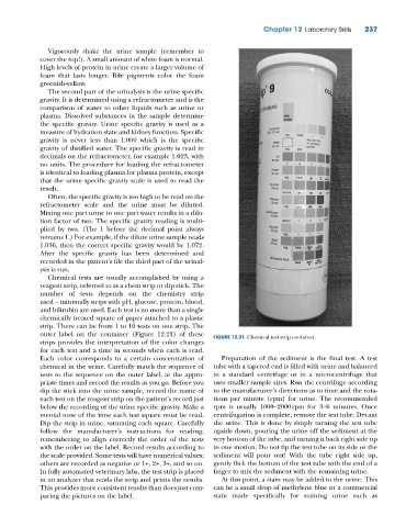
outer label on the container (Figure 12.21) of these FIGURE 12.21 Chemical test strip container.
strips provides the interpretation of the color changes
for each test and a time in seconds when each is read.
Each color corresponds to a certain concentration of Preparation of the sediment is the final test. A test
chemical in the urine. Carefully match the sequence of tube with a tapered end is filled with urine and balanced
tests to the sequence on the outer label, at the appro- in a standard centrifuge or in a microcentrifuge that
priate times and record the results as you go. Before you uses smaller sample sizes. Run the centrifuge according
dip the stick into the urine sample, record the name of to the manufacturer’s directions as to time and the rota-
each test on the reagent strip on the patient’s record just tions per minute (rpm) for urine. The recommended
below the recording of the urine specific gravity. Make a rpm is usually 1000–2000 rpm for 3–6 minutes. Once
mental note of the time each test square must be read. centrifugation is complete, remove the test tube. Decant
Dip the strip in urine, saturating each square. Carefully the urine. This is done by simply turning the test tube
follow the manufacturer’s instructions for reading, upside down, pouring the urine off the sediment at the
remembering to align correctly the order of the tests very bottom of the tube, and turning it back right side up
with the order on the label. Record results according to in one motion. Do not tip the test tube on its side or the
the scale provided. Some tests will have numerical values; sediment will pour out! With the tube right side up,
others are recorded as negative or 1+, 2+, 3+, and so on. gently flick the bottom of the test tube with the end of a
In fully automated veterinary labs, the test strip is placed finger to mix the sediment with the remaining urine.
in an analyzer that reads the strip and prints the results. At this point, a stain may be added to the urine. This
This provides more consistent results than does just com- can be a small drop of methylene blue or a commercial
paring the pictures on the label. stain made specifically for staining urine such as