
Page 248 - Zoo Animal Learning and Training
P. 248
232 Tasks for the Veterinary Assistant
blood doesn’t thin out enough and too fast it
streaks.
4. Failure to keep the spreader slide at a 45‐degree
angle. Too steep an angle or too shallow an angle
will not give the blood a chance to thin out to the
monolayer.
5. Failure to maintain constant pressure on the
spreader slide throughout the full length of the
slide. This usually results in streaks and or wobbles
in the smear.
6. Pulling, instead of pushing the spreader slide. If you
put the spreader slide into the blood the wrong way
you don’t get a good monolayer.
7. Not keeping the stains fresh and free of contami-
nates. Old stain or stain that has precipitated out
will leave small dark marks on the cells. This could
be mistaken for blood pathogens and an animal may
be treated for something they don’t have!
The veterinary technician examines the blood smear
under the oil immersion lens of the microscope. This
procedure is called the differential cell count. It is done
to determine the percentage of each white blood cell
type present, the morphology of all cells seen, and the
presence of any infectious organisms.
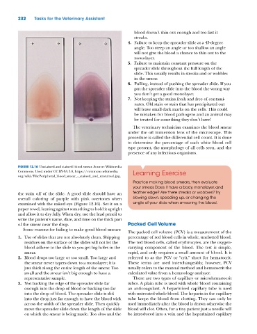
FIGURE 12.16 Unstained and stained blood smear. Source: Wikimedia
Commons. Used under CC BY‐SA 3.0, https://commons.wikimedia. Learning Exercise
org/wiki/File:Peripheral_blood_smear_‐_stained_and_unstained.jpg.
Practice making blood smears, then evaluate
your smear. Does it have a body, monolayer, and
the stain off of the slide. A good slide should have an feather edge? Are there streaks or wobbles? Try
overall coloring of purple with pink overtones when slowing down, speeding up, or changing the
examined with the naked eye (Figure 12.16). Set it on a angle of your slide when smearing the blood.
paper towel, leaning against something to hold it upright
and allow it to dry fully. When dry, use the lead pencil to
write the patient’s name, date, and time on the thick part
of the smear near the drop. Packed Cell Volume
Some reasons for failing to make good blood smears:
The packed cell volume (PCV) is a measurement of the
1. Use of slides that are not absolutely clean. Shipping percentage of red blood cells in whole, unclotted blood.
residues on the surface of the slides will not let the The red blood cells, called erythrocytes, are the oxygen‐
blood adhere to the slide so you get big holes in the carrying component of the blood. The test is simple,
smear. rapid, and only requires a small amount of blood. It is
2. Blood drops too large or too small. Too large and referred to as the PCV or “crit,” short for hematocrit.
the smear never tapers down to a monolayer; it is These terms are used interchangeably; however, PCV
just thick along the entire length of the smear. Too usually refers to the manual method and hematocrit the
small and the smear isn’t big enough to have a calculated value from a hematology analyzer.
representative sample. There are two types of capillary or microhematocrit
3. Not backing the edge of the spreader slide far tubes. A plain tube is used with whole blood containing
enough into the drop of blood or backing too far an anticoagulant. A heparinized capillary tube is used
into the drop of blood. The spreader slide is slid with untreated whole blood. The heparin in the capillary
into the drop just far enough to have the blood wick tube keeps the blood from clotting. They can only be
across the width of the spreader slide. Then quickly used immediately after the blood is drawn otherwise the
move the spreader slide down the length of the slide blood will clot. Often, for a tiny patient just a needle will
on which the smear is being made. Too slow and the be introduced into a vein and the heparinized capillary