
Page 250 - Zoo Animal Learning and Training
P. 250
234 Tasks for the Veterinary Assistant
results in g/dL (grams per 100 mL) into the lab log book
Line up on and in the patient’s file.
100 When finished, clean and disinfect the work area,
centrifuge, and refractometer, put materials away, and
Plasma discard blood contaminated materials in a biohazard
waste container.
Learning Exercise
Read line Buffy coat
for PCV Write out the procedures for PCV and plasma pro-
tein determination in your reference book.
Packed red
blood cells
Blood Chemistry and Electrolyte
Determinations
Line up on Clay seal
zero
In the past, blood chemistries and electrolyte determina-
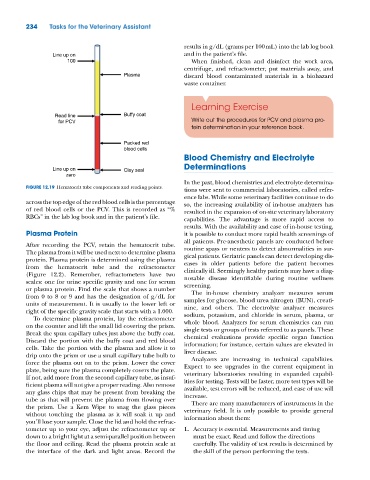
FIGURE 12.19 Hematocrit tube components and reading points.
tions were sent to commercial laboratories, called refer-
ence labs. While some veterinary facilities continue to do
across the top edge of the red blood cells is the percentage so, the increasing availability of in‐house analyzers has
of red blood cells or the PCV. This is recorded as “% resulted in the expansion of on‐site veterinary laboratory
RBCs” in the lab log book and in the patient’s file. capabilities. The advantage is more rapid access to
results. With the availability and ease of in‐house testing,
Plasma Protein it is possible to conduct more rapid health screenings of
all patients. Pre‐anesthetic panels are conducted before
After recording the PCV, retain the hematocrit tube. routine spays or neuters to detect abnormalities in sur-
The plasma from it will be used next to determine plasma gical patients. Geriatric panels can detect developing dis-
protein. Plasma protein is determined using the plasma eases in older patients before the patient becomes
from the hematocrit tube and the refractometer clinically ill. Seemingly healthy patients may have a diag-
(Figure 12.2). Remember, refractometers have two nosable disease identifiable during routine wellness
scales: one for urine specific gravity and one for serum screening.
or plasma protein. Find the scale that shows a number The in‐house chemistry analyzer measures serum
from 0 to 8 or 9 and has the designation of g/dL for samples for glucose, blood urea nitrogen (BUN), creati-
units of measurement. It is usually to the lower left or nine, and others. The electrolyte analyzer measures
right of the specific gravity scale that starts with a 1.000. sodium, potassium, and chloride in serum, plasma, or
To determine plasma protein, lay the refractometer
on the counter and lift the small lid covering the prism. whole blood. Analyzers for serum chemistries can run
single tests or groups of tests referred to as panels. These
Break the spun capillary tubes just above the buffy coat. chemical evaluations provide specific organ function
Discard the portion with the buffy coat and red blood information; for instance, certain values are elevated in
cells. Take the portion with the plasma and allow it to liver disease.
drip onto the prism or use a small capillary tube bulb to Analyzers are increasing in technical capabilities.
force the plasma out on to the prism. Lower the cover Expect to see upgrades in the current equipment in
plate, being sure the plasma completely covers the plate. veterinary laboratories resulting in expanded capabil-
If not, add more from the second capillary tube, as insuf- ities for testing. Tests will be faster, more test types will be
ficient plasma will not give a proper reading. Also remove available, test errors will be reduced, and ease of use will
any glass chips that may be present from breaking the increase.
tube as that will prevent the plasma from flowing over There are many manufacturers of instruments in the
the prism. Use a Kem Wipe to snag the glass pieces veterinary field. It is only possible to provide general
without touching the plasma as it will soak it up and information about them:
you’ll lose your sample. Close the lid and hold the refrac-
tometer up to your eye, adjust the refractometer up or 1. Accuracy is essential. Measurements and timing
down to a bright light at a semi‐parallel position between must be exact. Read and follow the directions
the floor and ceiling. Read the plasma protein scale at carefully. The validity of test results is determined by
the interface of the dark and light areas. Record the the skill of the person performing the tests.