Page 249 - Zoo Animal Learning and Training
P. 249
Chapter 12  Laboratory Skills  233


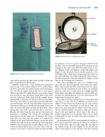
                                                                                                         Top lid


                                                                                                         Inside lid


                                                                                                         Slots for
                                                                                                         PCV tubes


                                                                FIGURE 12.18  Hematocrit centrifuge lids and slots.


                                                                the patient’s name on a piece of paper with the tube
                                                                location. This is especially important if running several
                                                                samples at the same time. Put the inside cover on the
                                                                tray (Figure 12.18) and tighten it down with either your
                                                                fingers or the lid plyers that is usually attached to the
             FIGURE 12.17  Hematocrit tubes and tray of clay sealant.  centrifuge with a chain. If you forget to put this cover on,
                                                                the tubes will spin out of the slots and be busted into a
                                                                fine bloody powder. Close the outside lid (Figure 12.18)
             tube will be put into the hub of the needle to draw out   and set the centrifuge for 5 minutes.
             just enough blood to fill the tube.                   Once the centrifuge has come to a complete stop,
               Supplies required for a PCV include two hematocrit   open the first and second lids and remove the tubes.
             tubes, a tray of clay sealant (Figure 12.17), a Kem Wipe,   Place them beside the PCV card reader and the refrac-
             the PCV card reader, the refractometer, and a lens paper.   tometer for the technician to read. If there are multiple
             Cap the blood sample and gently mix it once again.   patients, make sure to keep everyone’s sample separate.
             Then hold both PCV tubes between your index finger   Some clinics may allow the assistant to read the PCV and
             and thumb on your dominant hand. Slide both tubes   perform the plasma protein. If so, the following are the
             into the blood and hold them and the sample almost   instructions for these two tests.
             horizontally in order for the blood to flow into the tubes.   The tubes will have changed from a homogeneous
             Once they have filled to about three‐quarters full move   red color to one with three different colored layers
             your index finger over the top of the tubes to prevent the   (Figure 12.19). From the bottom up, the clay sealant,
             blood from flowing out. Set the blood sample down and   a dark red layer composed of red blood cells, just
             wipe the end of the tubes off with the Kem Wipe. Then   above that and often barely visible a white line is seen.
             plunge the ends of the tubes into the clay to seal them.   This is called the buffy coat and is composed of white
             Remove the tubes from the clay and put them into the   blood cells and platelets. Above that is the clear to
             hematocrit centrifuge. The hematocrit  centrifuge will   golden liquid which is the plasma. If the color is dark
             have slots to put the tubes into. To balance the centri-  yellow, pink or red, or white alert the technician or
             fuge, put one tube with the sealed end towards the   veterinarian.
             outside of the circle and then directly across the circle   To determine the PCV, a microhematocrit reader or
             from the first tube lay the second tube into the slot, again   PCV card is used. The card can be linear or circular in
             with the clay end toward the outside (Figure 12.18), oth-  form. With either type of reader, align the top edge of
             erwise the blood will be spun out of the tubes. Note the   the sealant on the zero line (Figure 12.19) and the top
             number on the slot in which the tubes are placed. Write   edge of the plasma on the 100 line. The line running

