Page 300 - Zoo Animal Learning and Training
P. 300
284    Tasks for the Veterinary Assistant


                                                          Occlusal


                                      Crown
                                                                                Coronal
                                    Enamel

                                                                          Attached
                                       Neck
                                                                           gingiva
                                                                         Sulcus
                                      Dentin
                                  Pulp chamber
                                   nerve/blood
                                       vessel
                                         Root                                    Apical

                                                      Furcation
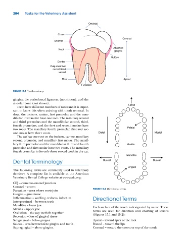
          FIGURE 15.1  Tooth anatomy.


          gingiva, the periodontal ligament (not shown), and the
          alveolar bone (not shown).                                                Labial
            Teeth have different numbers of roots and it is impor-
          tant to know this when assisting with tooth removal. In
          dogs, the incisors, canine, first premolar, and the man-
          dibular third molar have one root. The maxillary second
          and third premolars and the mandibular second, third,
          fourth premolars, and the first and second molars have
          two roots. The maxillary fourth premolar, first and sec-                 Palatal
          ond molar have three roots.                            Distal                              Mesial
            The cat has one root on the incisors, canine, maxillary
          second premolar, and maxillary first molar. The maxil-
          lary third premolar and the mandibular third and fourth                  Maxilla
          premolar, and first molar have two roots. The maxillary
          fourth premolar is the only three rooted teeth in the cat.
                                                                                   Mandible
          Dental Terminology                                        Buccal                          Buccal

                                                                                    Lingual
          The following terms are commonly used in veterinary
          dentistry. A complete list is available at the American
          Veterinary Dental College website at www.avdc.org:
          CEJ – cemento‐enamel junction
          Coronal – crown
          Furcation – area where roots join                  FIGURE 15.2  Directional terms.
          Gingiva – gum tissue
          Inflammation – swelling, redness, infection        Directional Terms
          Interproximal – between teeth
          Mandible – lower jaw                               Each surface of the tooth is designated by name. These
          Maxilla – upper jaw                                terms are used for direction and charting of lesions
          Occlusion – the way teeth fit together             (Figures 15.1 and 15.2):
          Recession – loss of gingival tissue
          Subgingival – below gingiva                        Apical – toward apex of the root
          Sulcus – area between free gingiva and tooth       Buccal – toward the lips
          Supragingival – above gingiva.                     Coronal – toward the crown or top of the tooth

