
Page 33 - REVISED GP Fall 2021 - ready for posting
P. 33
Principles of Odontogenic Infection Management
By Pooja Gangwani, DDS, MPH
Case Report Clinical exam: Laboratory findings:
Pertinent findings on his exam revealed His white blood count (WBC) at the time
Chief complaint: brawny, indurated, and painful neck swell- of admission was elevated at 13.5 with in-
A 63-year-old male presented to the emer- ing extending from the right chin to the left creased neutrophils at 12.8. His basic met-
gency department, referred by his general angle of mandible, with blunting of inferior abolic panel (BMP) revealed raised blood
dentist for evaluation of a large swelling in border of the mandible. There was erythema urea nitrogen (BUN) and creatinine levels
his neck. of the overlying skin. His mouth opening to 42 and 1.61, respectively. His blood glu-
was approximately 30 mm, and the floor cose levels were elevated to 253.
History of present illness: of his mouth was elevated and edematous
The patient was recently discharged from upon palpation. As per his dentist’s thor- Management:
the intensive care unit (ICU) of another ough examination, tooth # 20 was necrotic. The patient was taken to the operating room
hospital due to an improvement of his neck for incision and drainage of the multi-space
swelling after intravenous antibiotics. Six CT maxillofacial with contrast:
days later he was referred back to the emer- Imaging revealed multiloculated fluid col- infection. Aggressive surgical drainage and
gency room as his neck swelling did not lection with a thin enhancing wall and sur- debridement of bilateral submandibular,
resolve. (Figure 1) Furthermore, he was at rounding fat, collectively measuring 2.9 x submental and left sublingual spaces was
risk of airway compromise. Despite compli- 6.0 x 2.3 cm. There was no identifiable peri- performed. Cultures were obtained. Penrose
ance with Augmentin after his discharge, he apical dental lucency or cortical disruption. drains were placed in bilateral subman-
progressively developed persistent bilateral (Figures 2A and 2B) dibular and submental spaces and secured
with 3-0 silk suture. The source of the in-
fection was eliminated by extraction of
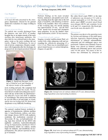
Figure 1. Significant fluctuant swell-
ing of bilateral neck and blunting of
the inferior border of the mandible.
neck swelling and pain. His symptoms had
been present for about two weeks. The pa-
tient did not recollect preceding pain associ- Figure 2A. Coronal view of contrast-enhanced CT scan, demonstrating
ated with any of his teeth. He reported hav- large hypodense area representing loculation.
ing subjective fevers and chills in addition
to odynophagia. The patient also stated that
he was not able to eat and drink, as it hurt,
and he was not feeling well. He denied any
dysphonia or any difficulty breathing.
Past medical history/Medications/Allergies:
The patient’s medical history included di-
agnoses of hypertension, diabetes mellitus
type 2, hyperlipidemia, asthma, and mini-
mal change disease (MCD) with nephrotic
syndrome. His medications consisted of
furosemide 40 mg, cyclophosphamide 50
mg, high dose steroids for his MCD with
nephrotic syndrome, hydrochlorothiazide
25 mg for hypertension, Symbicort 160-4.5
mcg/actuation inhaler for asthma, insulin
lispro for diabetes, and fenofibrate for hy-
perlipidemia. The patient reported an aller- Figure 2B. Axial view of contrast-enhanced CT scan, demonstrating
gy to lisinopril. large hypodense area representing loculation.
www.nysagd.org l Fall 2021 l GP 33