
Page 36 - REVISED GP Fall 2021 - ready for posting
P. 36
of transecting this vessel, the operator should be prepared to first apply The lateral wall of the right maxilla presented a smooth surgical site
direct pressure or attempt to burnish the vessel with a rotary drill. with the landmarks of teeth #2 and #4 easily visualized (Figure 4). A
Today we have a wide choice of osseous graft materials. 10,11 This now basket bur XRT084025 (Dentium, Engle-wood Cliffs, NJ) (Figure 5)
includes supplements such as the growth factors rhBMP and the was placed on a surgical handpiece with internal irrigation and at 500
12
patient’s own mesenchymal stem cells, which are harvested by spinning
down drawn blood. The choice is largely practitioner preference.
13
Case Report
In this case the loss of the first molar #3 has resulted in the pneumatization
of the sinus into the space normally occupied by the molar root system.
This creates a valley, which would normally be a fortunate anatomical
contour for any one of the sinus lift procedures (Figures 2,3). However,
after further inspection, the patient, an adult male, reported he had
an endodontically
treated tooth that was
symptomatic for years
and subsequently
under-went a
difficult extraction
to remove it. The 4 Figure 4. Dentium 5 Figure 5. DASK Basket
R extraction procedure Dentium Advanced Sinus Kit DASK DASK Basket bur XRT084025 on left
left the patient with Advanced Sinus Kit DASK. bur XRT084025 on left.
a highly irregular rpm was stroked across the bone in a feather brush motion. The bur was
ridge and variable held so that the rotational surface approached the bone in a downward
bone thickness. He direction. Copious water provided ample irrigation and allowed easy
presented for an visualization of the progress. A space one and a half times the diameter
implant consultation of the bur was reduced to a thin depressible veneer of bone. The bur was
2 Figure 2. Pre-op CBCT with inflamed to replace the missing turned perpendicular to the bone and the center water port directed to
sinus membrane, minimal bone height.
Pre-op CBCT with inflamed sinus membrane, minimal bone height tooth. the bone. A light brushing motion across the surface of the prepared bed
removed the remaining bone and the water jet in the center depressed
During the initial exam the the membrane away from the cutting edge of the basket bur (Figure 6)
mesial-distal space between #2
and #4 was 12mm, adequate
for an implant supported crown
and the ridge width, 8 mm, was
adequate to house an implant
with 2 mm of bone on each side.
However, the vertical bone height
from the crest of the ridge to the
floor of the sinus measured 3 mm
or less and was very irregular.
The patient was advised of the
minimums needed for predictable
implant placement and a lateral
Figure 3. Pre-op CBCT lateral entry sinus graft was suggested.
3
Pre-op CBCT lateral view, poor bone height, thickened sinus mucosa,
view, poor bone height, thick- Because of the irregular ridge 6Figure 6. Initial osteotomy. Figure 7. DASK hand
septa
Initial osteotomy
ened sinus mucosa, septa. contour a lateral approach was instruments XSE4L on left,
XSE3L in center.
preferred in this case. The patient 7 DASK hand instruments XSE4L on left, XSE3L in center
was also advised of the thickened sinus membrane and a referral to an The membrane was gently depressed with the hand instrument, XSE4L,
otolaryngologist was made. After a course of Amoxicillin and a waiting which has a mushroom shape (Figure 7) and allows for the membrane to
period of 3 months the sinus improved and the treatment was scheduled. be depressed and dissected from the inner surface of bone. The edge of
the instrument can be slipped between the inner surface of bone and the
The Surgical Procedure membrane. Once this dissection is started, the XSE3L or XSE1L can be
Anesthesia was obtained by blocking the secondary division of the used to continue the dissection in all directions around the osteotomy
maxillary nerve (V2). This was accomplished by infiltrating 1.5 ml. of (Figures 8.9). The key is to place the sharp end of the instrument on
Articaine hydrochloride 4% and epinephrine 1:100,000 (Septodont, St- the bone and allow the instrument to
Maur-des-Fossés, France) posterior to the right tuberosity and ½ inch pick the membrane off the inner surface
deep into the pterygomaxillary space. The V2 block is very effective
because it provides anesthesia for the lateral portion of the nose, cheek,
palate, maxillary teeth and sinus. To improve hemostasis, a supplementary
infiltration was made on the midline of the ridge and along the anticipated
incision lines on the buccal mucosa superior to teeth #2 and #4.
The patient was prepared in the usual manner and the surgical team
presented gloved and gowned. A #15 scalpel was used to make two
incisions in the vestibular mucosa, one superior to tooth #2 and the other
superior to tooth #4. A conservative 40 mm trapezoid access flap, from
the midline of #2 to the midline of #4, was planned to create room for a
20 mm osseous window. An incision was made on the crest of the ridge
extending to the bone and connected to the buccal incisions, creating a
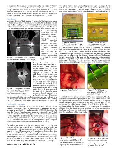
Initial placement of hand instrument XSE3L to dissect membrane from the Figure 9. XSE3L placed to
freely moving flap that extended to the zygoma. The gingiva and mucosa Figure 8. Initial placement of 9 XSE3L placed to the floor of the sinus and relieving the sinus membrane
8
internal aspect of the lateral sinus wall
were elevated with the periosteum in one continuous surgical flap. hand instrument XSE3L to dis- the floor of the sinus and
from the lateral wall
sect membrane from the internal relieving the sinus membrane
www.nysagd.org l Fall 2021 l GP 36 aspect of the lateral sinus wall. from the lateral wall.