
Page 35 - REVISED GP Fall 2021 - ready for posting
P. 35
Lateral Sinus Lift: A Case Report
By Joseph DiDonato, III, DDS, MBA, FAGD
The lateral wall maxillary sinus lift has been a predictable procedure to be filled with copious osseous graft. The raised membrane and the
for developing adequate bone in the posterior maxilla for decades. The lateral wall window can be supported with a collagen membrane and
sinus lift, whether it is accomplished through the lateral wall of the sinus then the entire flap closed.
(Tatum Lateral Access) or through the ridge (osteotome technique)
provides a means of increasing the recipient site for implants in an In some cases operators prefer to use ultrasonic tips that cut hard tissue
edentulous and atrophied posterior maxilla. but will leave the soft membrane intact. Alternatively, the use of a
rotary ‘basket’ can be used, as in the case presented, to remove the thin
The lateral wall procedure is a modified Caldwell-Luc procedure cortical plate and leave the membrane intact.
1
and was first orally reported by Tatum in the 1970’s, who began by
onlaying resorbed ridges with harvested autogenous rib bone. He And lastly, in cases where the anatomy is suitable, one can approach
2
found, however, that the graft decreased the intradental height and from the ridge. In this technique the operator drills to within a millimeter
resulted in very little bone for implant placement. He later decided to of the sinus floor and then breaks the remaining bone with a sized
enter the sinus to increase the bone height internally and not affect the osteotome. This lifts the sinus membrane and creates a space where
intradental space. He first lectured about the procedure to a study club bone graft can be injected through the osteotomy, and then the implant
in Alabama. The first report in the literature was by Boyne and James in can be placed. Alternatively, another technique is to drill through the
1980 followed by Tatum. The procedure as it is performed today has cortical plate on the floor and then to inject water to gently lift the
3,4
several variations including entering the sinus through the ridge. It is a membrane from its bed. In all cases having suitable instrumentation
procedure that has given many patients the benefits of implant therapy for manipulating the membrane makes the task much more manageable
who might otherwise be left wearing removable appliances. and provides a more predictable outcome.
The lateral wall of the sinus, bounded anteriorly by the cuspid and The treatment planning for the lateral procedure (Tatum Lateral
superiorly by the zygoma, is relatively thin, in many cases, one to two Window) is begun by obtaining a CBCT of the patient. The CBCT
5
millimeters. This affords a relatively easy and immediate access to the is studied to determine the anatomy of the antrum, specifically the
maxillary sinus directly superior to the maxillary posterior ridge. This thickness of the floor of the sinus in the vicinity of the intended implant
access allows the operator to lift the Schneiderian membrane and place placement. Misch and others recommend that if the antrum floor is
9
bone graft directly on the maxillary sinus floor. 5 mm or less, than a sinus lift/implant placement should be done as
a two-stage procedure, first the sinus lift, then wait 6 - 10 months for
The Schneiderian membrane has been described as a mucous membrane healing, then place the implant. With bone heights between 5 - 10 mm,
6
that covers the interior of the maxillary sinus (antrum). Histologically, the graft and the implant placement can be done at the same procedure.
it consists of the periosteum covered by a layer of pseudostratified The determining factors are, of course, initial implant stability and final
ciliated epithelium and highly vascularized connective tissue, which torque value used during placement.
contains mesenchymal stem cells that have the ability to form bone.
7
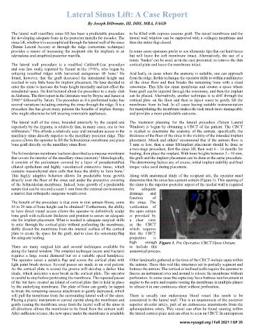
This highly adaptive behavior allows for predictable bone growth Along with anatomical study of the recipient site, the operator must
directly over the floor of the sinus and under the protective covering determine that the sinus has a patent ostium (Figure 1). This opening of
of the Schneiderian membrane. Indeed, bone growth of a predictable the sinus in the superior-posterior aspect of the medial wall is required
nature that can be secured a scant 1 mm from the external environment, for adequate
a marvel that orthopedic surgeons would covet. drainage and
function of
The benefit of the procedure is that even in thin antrum floors, some the sinus. The
16 to 20 mm of bone height can be obtained. Furthermore, the ability verification of
8
to have direct visual access allows the operator to definitively sculpt the open ostium R
bone graft with sufficient thickness and position to assure an adequate is provided by
site for implant placement. What is needed is adequate surgical skills a clear view
to enter through the cortical plate without perforating the membrane, in the CBCT
deftly dissect the membrane from the internal surface of the cortical which requires
plate to create the space for the graft, and to close the osteotomy/flap that the CBCT
for adequate healing. projection is
high enough Figure 1. Pre Operative CBCT Open Ostium.
There are many surgical kits and several techniques available for to include this
doing the lateral window. The simplest technique (score and fracture) anatomical process. 1 Pre Operative CBCT Open Ostium
requires a large round diamond bur on a variable speed handpiece.
The operator raises a suitable flap and scores the cortical plate with Other landmarks gathered at the time of the CBCT include septa within
light paint brush strokes. Several passes are made in an oval pattern. the antrum. These thin veil-like structures act to partially segment and
As the cortical plate is scored the groove will develop a darker blue buttress the antrum. The vertical or inclined walls require the operator to
shade, which indicates a near break in the cortical plate. The operator finesse an instrument over and around to release the membrane without
is careful to stop before perforating the membrane. The repeated passes perforation. In some cases the septa may have buttress reinforcement at
of the bur have created an island of cortical plate that is held in place angles to the septa and require teasing the membrane in multiple planes
by the underlying membrane. The plate of bone can gently be tapped to release it in one continuous sheet without perforation.
to break the remaining osseous attachment or gently depressed, which
will pull the membrane from the surrounding lateral wall of the sinus. There is usually one endosseous blood vessel that needs to be
Placing a plastic instrument or curved curette along the membrane and considered in the lateral wall. This is an anastomosis of the posterior
gently teasing the membrane away from the lateral wall of the sinus in superior alveolar artery, part of an endosseous anastomosis from the
all directions allows the membrane to be freed from the antrum wall. sphenopalatine artery. This vessel can often be found running within
After sufficient release, the new space under the membrane is available the lateral cortical plate and can often be seen on CBCT. In anticipation
www.nysagd.org l Fall 2021 l GP 35