
Page 9 - GP Spring 2020
P. 9
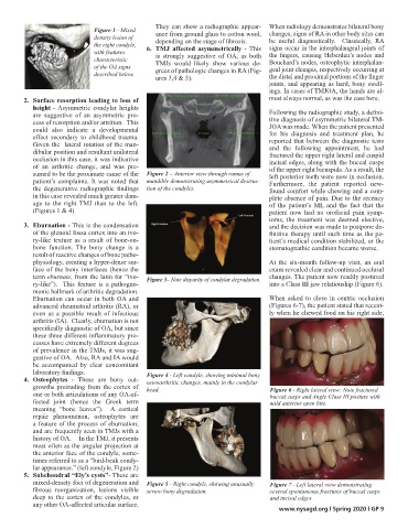
Figure 1 - Mixed     They can show a radiographic appear-  When radiology demonstrates bilateral bony
                               density lesion of      ance from ground glass to cotton wool,  changes, signs of RA in other body sites can
                               the right condyle,     depending on the stage of fibrosis.             be useful diagnostically.  Classically, RA
                               with features      6.  TMJ  affected  asymmetrically  - This  signs occur in the interphalangeal joints of
                               characteristic      is strongly suggestive of OA,  as both  the fingers, causing Heberden’s nodes and
                               of the OA signs      TMJs would  likely  show various  de-  Bouchard’s nodes, osteophytic interphalan-
                               described below.     grees of pathologic changes in RA (Fig-  geal joint changes, respectively occurring at
                                                ures 3,4 & 5).                    the distal and proximal portions of the finger
                                                                                  joints, and appearing as hard, bony swell-
                                                                                  ings. In cases of TMJOA, the hands are al-
        2.  Surface resorption leading to loss of                                 most always normal, as was the case here.
           height - Asymmetric  condylar heights
           are suggestive of an asymmetric  pro-                                  Following the radiographic study, a defini-
           cess of resorption and/or attrition.  This                             tive diagnosis of asymmetric bilateral TM-
           could  also  indicate  a  developmental                                JOA was made. When the patient presented
                                                                                  for his diagnosis and treatment plan, he
           effect  secondary to childhood  trauma.                                reported that between the diagnostic tests
           Given the  lateral rotation of the man-                                and the following appointment,  he had
           dibular position and resultant unilateral                              fractured the upper right lateral and cuspid
           occlusion in this case, it was indicative                              incisal edges, along with the buccal cusps
           of an arthritic  change, and was pre-                                  of the upper right bicuspids. As a result, the
           sumed to be the proximate cause of the   Figure 2 – Anterior view through ramus of   left posterior teeth were now in occlusion.
           patient’s complaints. It was noted that   mandible demonstrating asymmetrical destruc-  Furthermore,  the  patient  reported  new-
           the  degenerative  radiographic  findings   tion of the condyles.      found comfort while chewing and a com-
           in this case revealed much greater dam-                                plete absence of pain. Due to the recency
           age to the right  TMJ than to the left.                                of the patient’s MI, and the fact  that  the
           (Figures 1 & 4)                                                        patient  now had no orofacial  pain symp-
                                                                                  toms, the treatment was deemed elective,
        3.  Eburnation - This is the condensation                                 and the decision was made to postpone de-
           of the glenoid fossa cortex into an ivo-                               finitive therapy until such time as the pa-
           ry-like texture as a result of bone-on-                                tient’s medical condition stabilized, or the
           bone function.  The bony change is a                                   stomatognathic condition became worse.
           result of reactive changes of bone patho-
           physiology, creating a hyper-dense sur-                                At the six-month follow-up visit, an oral
           face of the bony interfaces (hence the                                 exam revealed clear and continued occlusal
           term eburnate, from the latin for “ivo-  Figure 3– Note disparity of condylar degradation.  changes. The patient now readily postured
           ry-like”).   This  feature  is  a  pathogno-                           into a Class III jaw relationship (Figure 6).
           monic hallmark of arthritic degradation.
           Eburnation  can  occur  in  both  OA and                               When asked to close in centric occlusion
           advanced rheumatoid arthritis (RA), or                                 (Figures 6-7), the patient stated that recent-
           even  as a  possible  result  of infectious                            ly when he chewed food on his right side,
           arthritis (IA).  Clearly, eburnation is not
           specifically diagnostic of OA, but since
           these three different inflammatory pro-
           cesses have extremely different degrees
           of prevalence in the TMJs, it was sug-
           gestive of OA.  Also, RA and IA would
           be  accompanied  by  clear  concomitant
           laboratory findings.              Figure 4 - Left condyle, showing minimal bony
        4.  Osteophytes  -  These are  bony out-  osteoarthritic changes, mainly in the condylar
           growths protruding from the cortex of   head.                          Figure 6 - Right lateral view: Note fractured
           one or both articulations of any OA-af-                                buccal cusps and Angle Class III posture with
           fected  joint  (hence  the  Greek  term                                mild anterior open bite.
           meaning  “bone leaves”).    A cortical
           repair phenomenon, osteophytes are
           a feature of the process of eburnation,
           and are frequently seen in TMJs with a
           history of OA.     In the TMJ, it presents
           most often as the angular projection at
           the anterior face of the condyle, some-
           times referred to as a “bird-beak condy-
           lar appearance.” (left condyle, Figure 2)
        5.  Subchondral “Ely’s cysts”- These are
           mixed-density foci of degeneration and   Figure 5 - Right condyle, showing unusually   Figure 7 - Left lateral view demonstrating
           fibrous  reorganization,  lesions  visible   severe bony degradation.  several spontaneous fractures of buccal cusps
           deep to the cortex of the condyles, or                                 and incisal edges.
           any other OA-affected articular surface.
                                                                                     www.nysagd.org l Spring 2020 l GP 9