
Page 22 - GP Fall 2019
P. 22
Treatment of Cystic Lesion Concurrent with Fully Impacted
Mesiodens at the Base of the Nasal Cavity: A Case Report
by Raid Sadda, DDS, MS, Cristina Navarro Lama, DDS, Juliana Gomez, DDS
Abstract of large maxillary cysts typically includes tist performed extractions of teeth: #2, 3, 4,
Background: This study emphasizes the non-surgical root canal therapy when local- 5, 11, 18, 30 and 31. Teeth #16 and 17 were
importance of thorough diagnostics and a ized and/or surgical treatment that may con- removed to address her symptoms, but the
multidisciplinary approach in surgical man- sist of enucleation, marsupialization and/ procedures and subsequent pain control did
agement of a large maxillary cystic lesion or decompression. 1,3,4,6,7 Management of the not resolve her pain. The patient moved to
with a mesiodens in the nasal floor. benign cyst of the maxilla in this case report the United States and presented to the emer-
included marsupialization, decompression, gency room for the anterior maxillary pain
Case Description: We will evaluate the pa- endodontic therapy and enucleation. Plan- and swelling. They drained the abscess and
tient and review treatment planning of the ning was of particular importance, however, placed a drain which temporarily relieved
case in this report. A panoramic radiograph because of a mesiodens present in the nasal her symptoms; but the pain came back af-
revealed a well-defined radiolucency of the floor that initially appeared to be in close ter two months. She presented again to the
anterior maxilla and computed tomography proximity to the lesion. Mesiodens are not emergency room where they referred her to
revealed a mesiodens posterior to the lesion uncommon dental abnormalities, occurring the Department of Oral and Maxillofacial
in the nasal floor. The treatment was staged in 0.09 to 2.5% of the general population. Surgery.
8,9
consisiting of marsupialization followed by About 75% of the time, mesiodens are im-
total excision. The mesiodens was not asso- pacted, however they often interfere with A panoramic radiograph was taken and a
ciated with the lesion and thus not removed. eruption of other teeth, and are known to be large well-defined radiolucency from max-
Literature evaluating marsupialization of associated with dentigerous cyst formation, illary right canine to upper left first premo-
maxillary cysts was reviewed and seven rel- and remain unerupted. Typically, they oc- lar can be appreciated (Figure 2A). There
10
evant articles were found. cur in the anterior premaxilla in close prox- is a radiopacity in the center of the lesion
imity to the dentition. They result in cyst on the panoramic, later to be identified as a
Practical Implications: The priority was formation 11% of the time. 10 fully impacted mesiodens. Maxillary central
resolution of symptoms of facial pain and and lateral incisors presented no response to
swelling. Due to careful diagnosis and con- Case Description pulp testing and root canal treatment was
servative treatment, the symptoms were A 37-year-old female presented to the NYU completed. Meanwhile, to evaluate the ex-
eliminated and the prognosis is promising. Department of Oral and Maxillofacial Sur- tent of the lesion, a cone-beam computed
gery with a chief complaint of severe pain tomography (CBCT) scan of the region was
Background in the upper lip, anterior teeth and face. She completed and revealed a fully impacted
Concurrence of an odontogenic cystic le- also reported recurrent swelling for over 10 mesiodens in the posterior nasal floor, sit-
sion and a supernumerary tooth in the max- years. Clinical findings included expansion ting on the hard palate. The radiology report
illa is rare in the literature. This case report of the premaxilla localized to the vestibule established that at its largest point, the cyst
1
describes unique management of a benign at the midline accompanied by deviation of measured 80 mm x 80 mm and was filled
cyst of the maxilla that possesses no causal maxillary labial frenulum, and moderate to with a homogenous fluid with density. The
relationship with a nearby mesiodens pres- severe proclination of all remaining ante- superior border of the cyst extended to the
ent in the nasal floor. rior maxillary teeth (Figure 1). The patient nasal floor near an impacted mesiodens,
denied significant medical or social history deep and superior to cyst; both entities inde-
Maxillary cystic lesions are often asymp- and reported no history of trauma. pendent of each other and unrelated (Figure
tomatic, sometimes presenting with swell- 2B). CBCT also revealed a small radicular
ing, and are usually diagnosed during rou- Relevant past interventions were conducted cyst at the upper right lateral incisor #7
tine radiologic examination. Treatment in her country of origin where the local den- (Figure 3A) that reduced in size after root
1-5
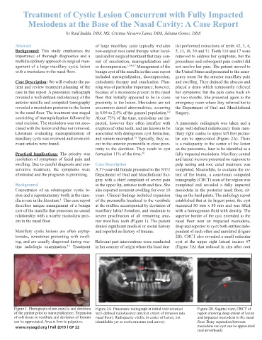
Figure 1. Photograph of pre-maxilla and dentition Figure 2A. Panoramic radiograph at initial visit revealed Figure 2B. Sagittal view, CBCT of
of the patient prior to marsupialization. Expansion well-defined radiolucency (unclear extent of invasion into region showing deep extent of lesion
of soft tissue in vestibule and deviation of frenum nasal floor). Radiopacity visible in center of lesion, not and impacted mesiodens in the nasal
can be appreciated. Area is firm to palpation. identifiable yet as tooth structure (red arrow). floor. Bony separation between
www.nysagd.org l Fall 2019 l GP 22 mesiodens and cyst can be appreciated
(red arrowhead).