
Page 23 - GP Fall 2019
P. 23
The bony defect where the cyst was re-
moved was assessed intraoperatively and
the supernumerary tooth was not visible or
palpable. It was determined that this presen-
tation in addition to the CBCT scan com-
pletely confirmed that the mesiodens was
not the cause of the cyst and removal of the
mesiodens would have caused more trauma
to the region. Specifically after the final his-
tologic diagnosis of benign cyst of maxilla
(Figure 5) it was proven that the mesiodens
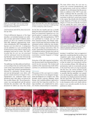
Figure 3A. Axial CBCT slice of lesion before Figure 3B. Axial CBCT slice after marsupialization
marsupialization. Blue arrow denoting small but prior to excision. New bone is visible growing was not causing the cystic lesion and ex-
radicular cyst treated by RCT. in from existing bony wall (red arrow).
canal treatment and will be observed at fol- for the first two months and once a month
low up visits. during the third and fourth months. The tube
drain was cleaned and irrigated at every visit
Clinical differential diagnosis of the large and the patient’s symptoms were evaluated.
maxillary cyst included residual cyst, radic- Four months after marsupialization, CBCT
ular cyst and, upon initial evaluation, denti- was performed to evaluate cyst size and
gerous cyst, due to its common association bone formation.(Figure 3B) Patient symp-
with fully impacted supernumerary teeth. toms of facial pain and facial swelling were
After evaluating the CBCT thoroughly, den- eliminated entirely within the four months. Figure 5. Histologic image of 100 x magnifica-
tigerous cyst was ruled out. A dentigerous Therefore, it was determined that the patient tion acquired from sample. Dark purple/black
cyst by definition is a cyst containing fluid was ready to undergo enucleation. After 16 structure indicates viable bone (red arrow),
localized between the reduced enamel of an weeks, enucleation was performed under lo- red arrowhead denotes marrow spaces.
unerupted tooth and its crown. The CBCT cal anesthesia and two samples were sent for
revealed that the cyst was not associated histologic evaluation. The histopathological tracting it would have been an aggressive
with the radiolucent lesion by demonstrat- examination revealed diagnosis of benign approach, considering that the excision was
ing that there was intact bone between the cyst of maxilla. completed under local anesthesia. There
cyst and the impacted supernumerary tooth was a possibility of injury to the floor of the
(Figure 2B). Extraction of the fully impacted mesiodens nose and creating an oro-nasal fistula, giv-
was discussed and it was determined that re- en that the palate was immediately inferior
The pathology was then addressed and treat- moval was not necessary. The location and to the mesiodens.(Figure 2B) Although su-
ed. According to the size and location of the condition of the supernumerary tooth will be pernumerary teeth do occasionally present
cystic lesion, surgical procedures conducted continue to be monitored at follow up visits. in the nasal floor and even erupt ectopically
were staged and included marsupialization into the nose, the curious position of this
followed by enucleation. Pre-op, intra-op Discussion mesiodens leads to some speculation. 8.9.11 It
and post-op photographs were taken with The purpose of this case report is to empha- is possible that the maxillary cyst extend-
patient consent and shown in Figure 4. Mar- size the importance of an adequate manage- ed so far through the midface such that the
supialization was conducted under local ment and diagnosis of a complex case. This mesiodens was displaced posteriorly further
anesthesia and a tube drain was placed for includes proper work-up, planning, initial into the nasal floor. This theory cannot be
four months (Figure 4B). The postoperative therapy and a multidisciplinary approach to confirmed without radiologic imaging prior
course was non-significant and the patient achieve the best long-term result for a be- to the formation of the cyst, which unfortu-
tolerated the procedure well. The patient nign maxillary cyst complicated by exten- nately is not currently possible for the de-
presented for follow up every two weeks sive borders and proximity to a mesiodens. partment to obtain.
Figure 4A. Closure obtained after first surgery Figure 4B. Intraoperative photo after drain re- Figure 4C. Patient in relaxed mouth position not
with drainage tube between maxillary central moval demonstrating residual defect in anterior showing drain and confirming that she is comfort-
incisors. Some reduction in expansion noted. buccal plate 16 weeks after marsupialization. able and satisfied with appearance.
Intraoperative image after enucleation of cyst.
www.nysagd.org l Fall 2019 l GP 23