
Page 49 - Libro vascular I
P. 49
Chap-04.qxd 29~8~04 13:21 Page 40
40
PERIPHERAL VASCULAR ULTRASOUND
When imaging tortuous vessels, it is useful to obtain images with the color box steered in differ- ent directions to visualize the blood flow along the entire vessel. If the direction of the blood flow changes in relation to the Doppler beam, a differ- ent Doppler frequency will be detected even though the blood velocity is the same. The color image will demonstrate a change of color within the vessel as the path of the vessel alters direction. Figure 4.7 shows an image of an internal carotid artery as its path dips deep into the neck. The arrows on the image show how the blood flow changes direction relative to the color Doppler beam, caus- ing a change in the Doppler frequency detected. This leads to a change in the color displayed, from red to orange and yellow then finally turquoise,
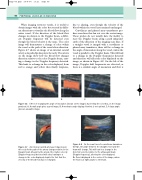
ABC
Figure 4.6 Effect of changing the angle of insonation (shown on the image), by steering the color box, on the image produced. A: A small angle gives a good image. B: A moderate angle displays flow but is not optimal. C: A large angle gives an unusable image.
due to aliasing, even though the velocity of the blood within the vessel has remained unchanged.
Curvilinear and phased array transducers pro- duce scan lines that fan out over the sector image. These probes do not usually have the facility to steer the Doppler beam along a path independ- ently of the direction of the imaging scan lines. If a straight vessel is imaged with a curvilinear or phased array transducer, there will be a change in the angle of insonation along the vessel, unless the vessel is parallel to the Doppler beam. This will lead to a change in the Doppler frequencies detected and therefore will affect the color displayed on the image, as shown in Figure 4.8. On the left of the image, Doppler shift frequencies are detected, as there is a suitable angle of insonation and flow is
Figure 4.7 An internal carotid artery as it dips deep in the neck. As the path of the artery changes relative to the Doppler beam (shown by the arrow), the relative velocity (Doppler frequency) detected will alter, leading to a change in the color displayed, despite the fact that the velocity of the blood flow has not changed.
Figure 4.8 As the scan lines of a curvilinear transducer diverge, the angle between the straight vessel and the beam will change. This will lead to a change in the detected velocity relative to the ultrasound beam (Doppler frequency), altering the color displayed.
No flow is displayed in the center of the image, where the flow is at right angles to the beam.
