
Page 43 - Basic _ Clinical Pharmacology ( PDFDrive )
P. 43
CHAPTER 2 Drug Receptors & Pharmacodynamics 29
Cytokine molecules
+ Cytokine
P~Y Y~P
R R R R
JAK JAK JAK JAK
Y~P
P~Y STAT STAT
Y~P
STAT STAT
P~Y
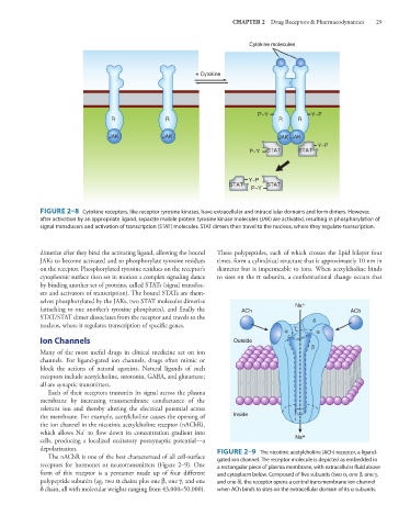
FIGURE 2–8 Cytokine receptors, like receptor tyrosine kinases, have extracellular and intracellular domains and form dimers. However,
after activation by an appropriate ligand, separate mobile protein tyrosine kinase molecules (JAK) are activated, resulting in phosphorylation of
signal transducers and activation of transcription (STAT) molecules. STAT dimers then travel to the nucleus, where they regulate transcription.
dimerize after they bind the activating ligand, allowing the bound These polypeptides, each of which crosses the lipid bilayer four
JAKs to become activated and to phosphorylate tyrosine residues times, form a cylindrical structure that is approximately 10 nm in
on the receptor. Phosphorylated tyrosine residues on the receptor’s diameter but is impermeable to ions. When acetylcholine binds
cytoplasmic surface then set in motion a complex signaling dance to sites on the α subunits, a conformational change occurs that
by binding another set of proteins, called STATs (signal transduc-
ers and activators of transcription). The bound STATs are them-
selves phosphorylated by the JAKs, two STAT molecules dimerize Na +
(attaching to one another’s tyrosine phosphates), and finally the ACh ACh
STAT/STAT dimer dissociates from the receptor and travels to the δ
nucleus, where it regulates transcription of specific genes. γ
α α
Ion Channels Outside
β
Many of the most useful drugs in clinical medicine act on ion
channels. For ligand-gated ion channels, drugs often mimic or
block the actions of natural agonists. Natural ligands of such
receptors include acetylcholine, serotonin, GABA, and glutamate;
all are synaptic transmitters.
Each of their receptors transmits its signal across the plasma
membrane by increasing transmembrane conductance of the
relevant ion and thereby altering the electrical potential across
the membrane. For example, acetylcholine causes the opening of Inside
the ion channel in the nicotinic acetylcholine receptor (nAChR),
+
which allows Na to flow down its concentration gradient into Na +
cells, producing a localized excitatory postsynaptic potential—a
depolarization. FIGURE 2–9 The nicotinic acetylcholine (ACh) receptor, a ligand-
The nAChR is one of the best characterized of all cell-surface gated ion channel. The receptor molecule is depicted as embedded in
receptors for hormones or neurotransmitters (Figure 2–9). One a rectangular piece of plasma membrane, with extracellular fluid above
form of this receptor is a pentamer made up of four different and cytoplasm below. Composed of five subunits (two α, one β, one γ,
polypeptide subunits (eg, two α chains plus one β, one γ, and one and one δ), the receptor opens a central transmembrane ion channel
δ chain, all with molecular weights ranging from 43,000–50,000). when ACh binds to sites on the extracellular domain of its α subunits.