
Page 44 - Basic _ Clinical Pharmacology ( PDFDrive )
P. 44
30 SECTION I Basic Principles
results in the transient opening of a central aqueous channel, concentration of the intracellular second messenger. For cAMP,
approximately 0.5 nm in diameter, through which sodium ions the effector enzyme is adenylyl cyclase, a membrane protein that
penetrate from the extracellular fluid to cause electrical depolar- converts intracellular adenosine triphosphate (ATP) to cAMP.
ization of the cell. The structural basis for activating other ligand- The corresponding G protein, G , stimulates adenylyl cyclase after
s
gated ion channels has been determined recently, and similar being activated by hormones and neurotransmitters that act via
general principles apply, but there are differences in key details specific G -coupled receptors. There are many examples of such
s
that may open new opportunities for drug action. For example, receptors, including α and β adrenoceptors, glucagon receptors,
receptors that mediate excitatory neurotransmission at central thyrotropin receptors, and certain subtypes of dopamine and
nervous system synapses bind glutamate, a major excitatory neu- serotonin receptors.
rotransmitter, through a large appendage domain that protrudes G and other G proteins activate their downstream effectors
s
from the receptor and has been called a “flytrap” because it physi- when bound by GTP and also have the ability to hydrolyze GTP
cally closes around the glutamate molecule; the glutamate-loaded (Figure 2–10); this hydrolysis reaction inactivates the G protein
flytrap domain then moves as a unit to control pore opening. but can occur at a relatively slow rate, effectively amplifying the
Drugs can regulate the activity of such glutamate receptors by transduced signal by allowing the activated (GTP-bound) G protein
binding to the flytrap domain, to surfaces on the membrane- to have a longer lifetime in the cell than the activated receptor
embedded portion around the pore, or within the pore itself. itself. For example, a neurotransmitter such as norepinephrine
The time elapsed between the binding of the agonist to a may encounter its membrane receptor for only a few milliseconds.
ligand-gated channel and the cellular response can often be mea- When the encounter generates a GTP-bound G molecule, how-
s
sured in milliseconds. The rapidity of this signaling mechanism is ever, the duration of activation of adenylyl cyclase depends on the
crucially important for moment-to-moment transfer of informa- longevity of GTP binding to G rather than on the duration of
s
tion across synapses. Ligand-gated ion channels can be regulated norepinephrine’s binding to the receptor. Indeed, like other
by multiple mechanisms, including phosphorylation and endocy- G proteins, GTP-bound G may remain active for tens of seconds,
s
tosis. In the central nervous system, these mechanisms contribute enormously amplifying the original signal. This mechanism also
to synaptic plasticity involved in learning and memory. helps explain how signaling by G proteins produces the phenom-
Voltage-gated ion channels do not bind neurotransmitters enon of spare receptors. The family of G proteins contains several
directly but are controlled by membrane potential; such channels functionally diverse subfamilies (Table 2–1), each of which medi-
are also important drug targets. Drugs that regulate voltage-gated ates effects of a particular set of receptors to a distinctive group
channels typically bind to a site of the receptor different from of effectors. Note that an endogenous ligand (eg, norepinephrine,
the charged amino acids that constitute the “voltage sensor” acetylcholine, serotonin, many others not listed in Table 2–1)
domain of the protein used for channel opening by membrane may bind and stimulate receptors that couple to different subsets
potential. For example, verapamil binds to a region in the pore of
voltage-gated calcium channels that are present in the heart and
in vascular smooth muscle, inhibiting the ion conductance sepa- Agonist
rately from the voltage sensor, producing antiarrhythmic effects,
and reducing blood pressure without mimicking or antagonizing
any known endogenous transmitter. Other channels, such as the
CFTR, although not strongly sensitive to either a known natural R * Cell membrane
ligand or voltage, are still important drug targets. Lumacaftor R
binds CFTR and promotes its delivery to the plasma membrane GTP
after biosynthesis. Ivacaftor binds to a different site and enhances GDP E
channel conductance. Both drugs act as allosteric modulators of
the CFTR and were recently approved for treatment of cystic G–GDP G–GTP
fibrosis, but each has a different effect.
E *
G Proteins & Second Messengers
P i
Many extracellular ligands act by increasing the intracellular con-
centrations of second messengers such as cyclic adenosine-3′,5′-
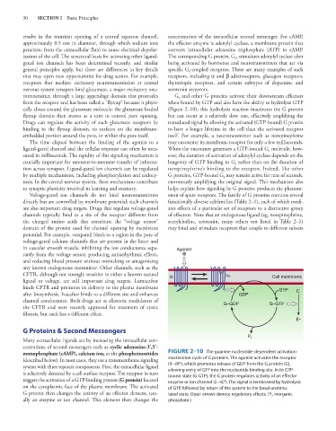
monophosphate (cAMP), calcium ion, or the phosphoinositides FIGURE 2–10 The guanine nucleotide-dependent activation-
(described below). In most cases, they use a transmembrane signaling inactivation cycle of G proteins. The agonist activates the receptor
system with three separate components. First, the extracellular ligand (R→R*), which promotes release of GDP from the G protein (G),
allowing entry of GTP into the nucleotide binding site. In its GTP-
is selectively detected by a cell-surface receptor. The receptor in turn bound state (G-GTP), the G protein regulates activity of an effector
triggers the activation of a GTP-binding protein (G protein) located enzyme or ion channel (E→E*). The signal is terminated by hydrolysis
on the cytoplasmic face of the plasma membrane. The activated of GTP, followed by return of the system to the basal unstimu-
G protein then changes the activity of an effector element, usu- lated state. Open arrows denote regulatory effects. (P i , inorganic
ally an enzyme or ion channel. This element then changes the phosphate.)