
Page 45 - Basic _ Clinical Pharmacology ( PDFDrive )
P. 45
CHAPTER 2 Drug Receptors & Pharmacodynamics 31
TABLE 2–1 G proteins and their receptors and effectors.
G Protein Receptors for Effector/Signaling Pathway
β-Adrenergic amines, histamine, serotonin, glucagon, and many ↑ Adenylyl cyclase →↑ cAMP
G s
other hormones
G i1 , G i2 , G i3 α 2 -Adrenergic amines, acetylcholine (muscarinic), opioids, Several, including:
serotonin, and many others ↓ Adenylyl cyclase →↓ cAMP
+
Open cardiac K channels →↓ heart rate
Odorants (olfactory epithelium) ↑ Adenylyl cyclase →↑ cAMP
G olf
Neurotransmitters in brain (not yet specifically identified) Not yet clear
G o
G q Acetylcholine (muscarinic), bombesin, serotonin (5-HT 2 ), and ↑ Phospholipase C →↑ IP 3 , diacylglycerol, cytoplasmic Ca 2+
many others
G t1 , G t2 Photons (rhodopsin and color opsins in retinal rod and ↑ cGMP phosphodiesterase →↓ cGMP (phototransduction)
cone cells)
cAMP, cyclic adenosine monophosphate; cGMP, cyclic guanosine monophosphate; IP3, inositol-1,4,5-trisphosphate.
of G proteins. The apparent promiscuity of such a ligand allows both proteins, allowing agonist binding to the receptor to effec-
it to elicit different G protein-dependent responses in different tively “drive” a nucleotide exchange reaction that “switches” the
cells. For instance, the body responds to danger by using catechol- G protein from its inactive (GDP-bound) to active (GTP-bound)
amines (norepinephrine and epinephrine) both to increase heart form. Figure 2–11 shows the main components schematically.
rate and to induce constriction of blood vessels in the skin, by
acting on G -coupled β adrenoceptors and G -coupled α adreno-
s
q
1
ceptors, respectively. Ligand promiscuity also offers opportunities
in drug development (see Receptor Classes & Drug Development Agonist
in the following text).
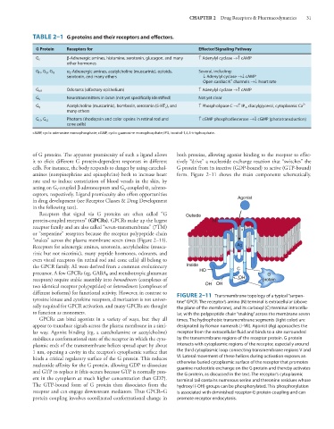
Receptors that signal via G proteins are often called “G Outside
protein-coupled receptors” (GPCRs). GPCRs make up the largest N
receptor family and are also called “seven-transmembrane” (7TM) II
or “serpentine” receptors because the receptor polypeptide chain I
“snakes” across the plasma membrane seven times (Figure 2–11). III
Receptors for adrenergic amines, serotonin, acetylcholine (musca- VII Ag IV
rinic but not nicotinic), many peptide hormones, odorants, and VI V
even visual receptors (in retinal rod and cone cells) all belong to
the GPCR family. All were derived from a common evolutionary Inside C
precursor. A few GPCRs (eg, GABA and metabotropic glutamate HO G
B
receptors) require stable assembly into homodimers (complexes of OH protein
two identical receptor polypeptides) or heterodimers (complexes of OH OH
different isoforms) for functional activity. However, in contrast to FIGURE 2–11 Transmembrane topology of a typical “serpen-
tyrosine kinase and cytokine receptors, dimerization is not univer- tine” GPCR. The receptor’s amino (N) terminal is extracellular (above
sally required for GPCR activation, and many GPCRs are thought the plane of the membrane), and its carboxyl (C) terminal intracellu-
to function as monomers. lar, with the polypeptide chain “snaking” across the membrane seven
GPCRs can bind agonists in a variety of ways, but they all times. The hydrophobic transmembrane segments (light color) are
appear to transduce signals across the plasma membrane in a simi- designated by Roman numerals (I–VII). Agonist (Ag) approaches the
lar way. Agonist binding (eg, a catecholamine or acetylcholine) receptor from the extracellular fluid and binds to a site surrounded
stabilizes a conformational state of the receptor in which the cyto- by the transmembrane regions of the receptor protein. G protein
plasmic ends of the transmembrane helices spread apart by about interacts with cytoplasmic regions of the receptor, especially around
1 nm, opening a cavity in the receptor’s cytoplasmic surface that the third cytoplasmic loop connecting transmembrane regions V and
binds a critical regulatory surface of the G protein. This reduces VI. Lateral movement of these helices during activation exposes an
nucleotide affinity for the G protein, allowing GDP to dissociate otherwise buried cytoplasmic surface of the receptor that promotes
guanine nucleotide exchange on the G protein and thereby activates
and GTP to replace it (this occurs because GTP is normally pres- the G protein, as discussed in the text. The receptor’s cytoplasmic
ent in the cytoplasm at much higher concentration than GDP). terminal tail contains numerous serine and threonine residues whose
The GTP-bound form of G protein then dissociates from the hydroxyl (-OH) groups can be phosphorylated. This phosphorylation
receptor and can engage downstream mediators. Thus GPCR–G is associated with diminished receptor-G protein coupling and can
protein coupling involves coordinated conformational change in promote receptor endocytosis.