
Page 47 - Basic _ Clinical Pharmacology ( PDFDrive )
P. 47
CHAPTER 2 Drug Receptors & Pharmacodynamics 33
A Agonist
Response
(cAMP)
1 2 3 4 5 1 2
Time
B Agonist in extracellular space
1 2
-OH -OH GRK
-OH -OH ATP
-OH -OH P Coated pit
P P
G S
5 β−Arr
3
4
P'ase 6 Lysosome
-OH P
-OH P
-OH Endosomes
P
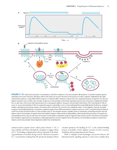
FIGURE 2–12 Rapid desensitization, resensitization, and down-regulation of β adrenoceptors. A: Response to a β-adrenoceptor agonist
(ordinate) versus time (abscissa). (Numbers refer to the phases of receptor function in B.) Exposure of cells to agonist (indicated by the light-
colored bar) produces a cyclic AMP (cAMP) response. A reduced cAMP response is observed in the continued presence of agonist; this “desensi-
tization” typically occurs within a few minutes. If agonist is removed after a short time (typically several to tens of minutes, indicated by broken
line on abscissa), cells recover full responsiveness to a subsequent addition of agonist (second light-colored bar). This “resensitization” fails to
occur, or occurs incompletely, if cells are exposed to agonist repeatedly or over a more prolonged time period. B: Agonist binding to receptors
initiates signaling by promoting receptor interaction with G proteins (G s ) located in the cytoplasm (step 1 in the diagram). Agonist-activated
receptors are phosphorylated by a G protein-coupled receptor kinase (GRK), preventing receptor interaction with G s and promoting binding of
a different protein, β-arrestin (β-Arr), to the receptor (step 2). The receptor-arrestin complex binds to coated pits, promoting receptor internal-
ization (step 3). Dissociation of agonist from internalized receptors reduces β-Arr binding affinity, allowing dephosphorylation of receptors by
a phosphatase (P’ase, step 4) and return of receptors to the plasma membrane (step 5); together, these events result in the efficient resensitiza-
tion of cellular responsiveness. Repeated or prolonged exposure of cells to agonist favors the delivery of internalized receptors to lysosomes
(step 6), promoting receptor down-regulation rather than resensitization.
2+
calcium-sensitive protein kinase called protein kinase C. IP is channels promotes the binding of Ca to the calcium-binding
3
water-soluble and diffuses through the cytoplasm to trigger release protein calmodulin, which regulates activities of other enzymes,
2+
of Ca by binding to ligand-gated calcium channels in the limit- including calcium-dependent protein kinases.
ing membranes of internal storage vesicles. Elevated cytoplasmic With its multiple second messengers and protein kinases, the
2+
Ca concentration resulting from IP -promoted opening of these phosphoinositide signaling pathway is much more complex than
3