
Page 102 - parasitology for medical and clinical laboratoryprofessionals
P. 102
82 CHAPTER 4
TABLE 4-1 Characteristics of Leishmanial Forms
OPISTHOMAS- TRYPOMAS-
AMASTIGOTE PROMASTIGOTE EPIMASTIGOTE TIGOTE TIGOTE
Size 2–5 μm × 1–3 μm 9–15 μm 9–15 μm Not available 12–35 μm
Flagella Reduced or Yes Yes Yes Yes
Presence absent
Shape Round (oval) Slender and Long, slightly Spindle-shaped Spindle-shaped,
elongated wider than C-shapes often
promastigotes seen in blood
films
lesions. For victims of leishmaniasis, recovery is based Disease Transmission
on cellular immunity where macrophages clear the in-
fectious sites of cellular debris and organisms. But those This disease is transmitted into the skin by sand flies and
with impaired immunity may progress to a more serious promastigotes of the three subspecies of L. tropica and
condition called diffuse cutaneous leishmaniasis, where are ingested by macrophages at the site of the injection of
nodular lesions occur particularly on the face, arms, and the promastigotes. Inside the macrophages the promas-
legs. In this case, the lesions are filled with parasites and tigotes progress into amastigote forms by multiplying
do not heal as quickly as uncomplicated cases. greatly. The host cell then ruptures and amastigotes are
free to enter uninfected macrophage cells. This continu-
ous cycle of invasion, multiplication, and cell rupture re-
Life Cycle
sults in the lesions and destruction of tissue.
The life cycle of Leishmania is no more complex than
that of other prominent parasites. The incubation pe- Laboratory Diagnosis
riod may range from a few weeks to three years depend-
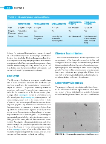
ing on the species. L. major has a more rapid onset of The presence of amastigotes is the definitive diagno-
symptoms and signs. Two morphologic stages occur in sis for leishmaniasis where aspirates from lesion sites
order for the transmission of the disease to take place. or a biopsy of tissue materials from an active lesion are
A stage called amastigotes occurs in a vertebrate host; stained with Wright’s or Giemsa stain, or a combination
after amastigotes, the promastigotes, a developmental and
infective stage, occurs only in the insect vector, so both
a host and a vector are required in order to transmit the
organism (Figure 4-4). As the vector bites the infected
host, amastigotes in macrophages (tissue cells that clean
up infections and debris) are ingested by the fly or other
vector, where the amastigotes undergo changes to become
flagellated forms called promastigotes. The promastigotes
then multiply rapidly before infecting the proboscis, or Source: Centers for Disease Control and Prevention (CDC)
biting parts of the vector, which is then transmitted to new
victims. Amastigotes often remain in the skin and other
tissues through the lymphatic system in the cutaneous
forms of the disease. In visceral forms, amastigotes migrate
within monocytes, types of protective white blood cells,
where the organisms migrate to the spleen, liver, and bone
marrow, and then continue to divide and multiply. FIGURE 4-4 Presence of promastigotes of Leishmania sp.