
Page 107 - parasitology for medical and clinical laboratoryprofessionals
P. 107
Blood (Intracellular) and Other Tissue Protozoa 87
blood samples is necessary to successfully demonstrate (Cox, 2002). Manson then turned his attention to bird
the presence of the organisms. Blood drawn just fol- malaria, where the fowl had contracted a species that
lowing the onset of a paroxysm (sudden recurrence of does not infect man, that of P. relictum. Culex is a genus
symptoms) such as a rapidly rising temperature should of mosquito where several species of this genus are vec-
contain free merozoites, which if visible do not provide tors of important diseases that greatly affect man, such
a means for identification of the species. The greatest as West Nile virus, filariasis, Japanese encephalitis,
number of parasites is present toward the end of a febrile St. Louis encephalitis, as well as avian malaria, a strain
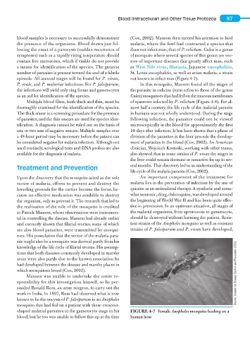
episode. All asexual stages will be found for P. vivax, not known to infect man (Figure 4-7).
P. ovale, and P. malariae infections. For P. falciparum, In this mosquito, Manson found all the stages of
the infections will yield only ring forms and gametocytes the parasite in culicine (term refers to those of the genus
as an aid for identification of the species. Culex) mosquitoes that had fed on the mucous membranes
Multiple blood films, both thick and thin, must be of sparrows infected by P. relictum (Figure 4-8). For al-
thoroughly examined for the identification of the species. most half a century, the life cycle of the malarial parasite
The thick smear is a screening procedure for the presence in humans was not wholly understood. During the stage
of parasites, and the thin smears are used for species iden- following infection, the parasites could not be viewed
tification. A diagnosis cannot be ruled out on the basis of microscopically in the blood for approximately the first
one or two sets of negative smears. Multiple samples over 10 days after infection. It has been shown that a phase of
a 48-hour period may be necessary before the patient can division of the parasites in the liver precede the develop-
be considered negative for malaria infection. Although not ment of parasites in the blood (Cox, 2002). An American
used routinely, serological tests and DNA probes are also clinician, Wojciech Krotoski, working with other teams,
available for the diagnosis of malaria. also showed that in some strains of P. vivax the stages in
the liver could remain dormant or nonactive for up to sev-
eral months. This discovery led to an understanding of the
Treatment and Prevention
life cycle of the malaria parasite (Cox, 2002).
Upon the discovery that the mosquito acted as the sole An important component of the treatment for
vector of malaria, efforts to prevent and destroy the malaria lies in the prevention of infection by the use of
breeding grounds for the carrier became the focus, be- quinine as an antimalarial therapy. A synthetic and some-
cause no effective medication was available to destroy what nontoxic, drug, chloroquine, was developed around
the organism, only to prevent it. The research that led to the beginning of World War II and has been quite effec-
the realization of the role of the mosquito is credited tive in prevention. In an optimum situation, all stages of
to Patrick Manson, whose observations were instrumen- the malarial organisms, from sprorozoite to gametocyte,
tal in controlling the disease. Manson had already earlier should be destroyed without harming the patient. Resis-
and correctly shown that filarial worms, some of which tant strains of the Anopheles mosquito as well as resistant
are also blood parasites, were transmitted by mosqui- strains of P. falciparum and P. vivax have developed,
toes. His postulation that the vector of the malaria para-
site might also be a mosquito was derived partly from his
knowledge of the life cycle of filarial worms. His assump-
tions that both diseases commonly developed in marshy
areas were also partly due to the known association he
had developed between the disease and marshy places in
which mosquitoes breed (Cox, 2002). Source: Centers for Disease Control and Prevention (CDC)
Manson was unable to undertake the entire re-
sponsibility for this investigation himself, so he per-
suaded Ronald Ross, an army surgeon, to carry out the
work in India. In 1897, Ross had observed what is now
known to be the oocysts of P. falciparum in an Anopheles
mosquito that had fed on a patient with these crescent-
shaped malarial parasites in the gametocyte stage in his FIGURE 4-7 Female Anopheles mosquito feeding on a
blood, but he too was unable to follow this up at the time human host