
Page 111 - parasitology for medical and clinical laboratoryprofessionals
P. 111
Blood (Intracellular) and Other Tissue Protozoa 91
Laboratory Diagnosis antigen detection tests (direct tests) are available. These
tests are not routinely performed in a clinical laboratory,
Most T. gondii infections are not diagnosed, because in- and are most often referred to a reference laboratory
dividuals who are exposed never feel ill or become symp- which specializes in these techniques.
tomatic or through a mistaken belief that a febrile disease
such as a cold or flu as an innocuous viral infection. Treatment and Prevention
Serological testing for IgM antibodies against T. gondii
is currently the most commonly employed diagnostic test Prevention is accomplished through proper personal
for acute infections. Convalescent IgG antibodies will oc- hygiene and avoiding the feces of cats and certain other
cur late in the infection after the acute phase has passed. animals such as pigs and sheep. Meat should be properly
Stained histological slides may reveal trophozoites and cooked before eating to ensure that the organisms are
cysts containing bradyzoites from surgically obtained tis- destroyed. It might be added that a great deal of expo-
sue biopsy samples. In the immunocompromised patient sure occurs in daily life, as up to one-third of the world’s
the presence of parasites in bronchoalveolar lavage or ce- population shows antibodies against the organism.
rebrospinal fluid (CSF) from a spinal tap may be used
effectively. Indirect testing for antibodies by an indirect
fluorescent antibody test (IFA), a Sabin-Feldman dye test, TRYPANOSOMES AND SLEEPING
indirect hemagglutination test, complement fixation, and
SICKNESS
Trypanosoma cruzi, Trypanosoma brucei gambiense, and
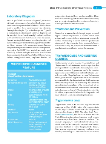
MICROSCOPIC DIAGNOSTIC
Trypanosoma brucei rhodensiense are three organisms that
FEATURE are responsible for several similar diseases by these blood-
and-tissue parasites. Trypanosoma cruzi is the causative
agent in the United States and parts of South and Cen-
General tral America for Chagas’s disease, whereas Trypanosoma
Classification—Sporozoan
brucei gambiense and Trypanosoma brucei rhodensiense
Organism Toxoplasma gondii are responsible for the disease in Africa. A different group
Specimen Required Generally by tissue of vectors is responsible for transmitting this disease in
biopsy specimens, each of the two hemispheres of the world, which will be
cerebrospinal fluid discussed later in this section. These related diseases are
Stage Trophozoites or cysts indeed serious, and the WHO estimates that up to16 to
are diagnostic 18 million people may be infected with this parasite and
Size Cysts measure from many others are at risk for contracting the disease.
12–100 μm
Oocysts in feces and Trypanosoma cruzi
soil range from 9–14 μm
Shape Oocysts are some- Trypanosoma cruzi is the causative organism for the
what oval American or New World variety of trypanosomiasis
Motility None called Chagas’s disease, named for Carlos Chagas who
Nucleus(i) Long, spherical discovered the disease in 1909. Also called South Amer-
nucleus at one end ican trypanosomiasis, the disease is found from the
of trophozoite United States to as far south as Argentina, which extends
Cytoplasm Smooth south to the tip of the South American continent. The
Other Features Tissue cysts contain causative organism, T. cruzi, infects both wild and do-
large numbers of mestic animals which may gain contact with each other,
slow-growing and makes it difficult to control the spread of the disease.
trophozoites Poor personal hygiene and unsanitary conditions com-
bine to place many poor farmers who handle animals