
Page 113 - parasitology for medical and clinical laboratoryprofessionals
P. 113
Blood (Intracellular) and Other Tissue Protozoa 93
Source: Centers for Disease Control and Prevention (CDC)
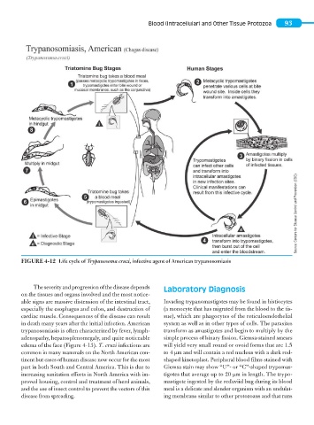
FIGURE 4-12 Life cycle of Trypanosoma cruzi, infective agent of American trypanosomiasis
The severity and progression of the disease depends Laboratory Diagnosis
on the tissues and organs involved and the most notice-
able signs are massive distension of the intestinal tract, Invading trypanomastigotes may be found in histiocytes
especially the esophagus and colon, and destruction of (a monocyte that has migrated from the blood to the tis-
cardiac muscle. Consequences of the disease can result sue), which are phagocytes of the reticuloendothelial
in death many years after the initial infection. American system as well as in other types of cells. The parasites
trypanosomiasis is often characterized by fever, lymph- transform as amastigotes and begin to multiply by the
adenopathy, hepatosplenomegaly, and quite noticeable simple process of binary fission. Giemsa-stained smears
edema of the face (Figure 4-13). T. cruzi infections are will yield very small round or ovoid forms that are 1.5
common in many mammals on the North American con- to 4 μm and will contain a red nucleus with a dark rod-
tinent but cases of human disease now occur for the most shaped kinetoplast. Peripheral blood films stained with
part in both South and Central America. This is due to Giemsa stain may show “U”- or “C”-shaped trypomas-
increasing sanitation efforts in North America with im- tigotes that average up to 20 μm in length. The trypo-
proved housing, control and treatment of herd animals, mastigote ingested by the reduviid bug during its blood
and the use of insect control to prevent the vectors of this meal is a delicate and slender organism with an undulat-
disease from spreading. ing membrane similar to other protozoans and that runs