
Page 133 - parasitology for medical and clinical laboratoryprofessionals
P. 133
Blood (Intracellular) and Other Tissue Protozoa 113
Symptoms
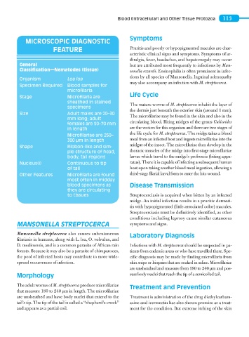
MICROSCOPIC DIAGNOSTIC
FEATURE Pruritis and poorly or hypopigmented macules are char-
acteristic clinical signs and symptoms. Symptoms of ar-
thralgia, fever, headaches, and hepatomegaly may occur
General but are attributed most frequently to infections by Man-
Classification—Nematodes (tissue) sonella ozzardi. Eosinophilia is often prominent in infec-
tions by all species of Mansonella. Inguinal adenopathy
Organism Loa loa
may also accompany an infection with M. streptocerca.
Specimen Required Blood samples for
microfilaria
Life Cycle
Stage Microfilaria are
sheathed in stained The mature worms of M. streptocerca inhabit the layer of
specimens
the dermis just beneath the exterior skin (around 1 mm).
Size Adult males are 20–30 The microfilariae may be found in the skin and also in the
mm long; adult
females are 50–70 mm circulating blood. Biting midges of the genus Culicoides
in length are the vectors for this organism and there are two stages of
the life cycle for M. streptocerca. The midge takes a blood
Microfilariae are 250–
300 μm in length meal from an infected host and ingests microfilariae into the
midgut of the insect. The microfilariae then develop in the
Shape Ribbon-like and sim-
ple structure of head, thoracic muscles of the midge into first-stage microfilariae
body, tail regions larvae which travel to the midge’s proboscis (biting appa-
Nucleus(i) Continuous to tip ratus). There it is capable of infecting a subsequent human
of tail host upon taking another blood meal ingestion, allowing a
Other Features Microfilaria are found third-stage filarial larval form to enter the bite wound.
most often in midday
blood specimens as Disease Transmission
they are circulating
to tissues Streptocerciasis is acquired when bitten by an infected
midge. An initial infection results in a pruritic dermati-
tis with hypopigmented (little associated color) macules.
Streptocerciasis must be definitively identified, as other
conditions including leprosy cause similar cutaneous
MANSONELLA STREPTOCERCA symptoms and signs.
Mansonella streptocerca also causes subcutaneous Laboratory Diagnosis
filariasis in humans, along with L. loa, O. volvulus, and
D. medinensis, and is a common parasite of African rain Infections with M. streptocerca should be suspected in pa-
forests. Because it may also be a parasite of chimpanzees, tients from endemic areas or who have travelled there. Spe-
the pool of infected hosts may contribute to more wide- cific diagnosis may be made by finding microfilaria from
spread occurrences of infection. skin snips or biopsies that are soaked in saline. Microfilariae
are unsheathed and measure from 180 to 240 μm and pos-
Morphology sess body nuclei that reach the tip of a semicoiled tail.
The adult worms of M. streptocerca produce microfilariae Treatment and Prevention
that measure 180 to 240 μm in length. The microfilariae
are unsheathed and have body nuclei that extend to the Treatment is administration of the drug diethylcarbam-
tail’s tip. The tip of the tail is called a “shepherd’s crook” azine and ivermectin has also shown promise as a treat-
and appears as a partial coil. ment for the condition. But extreme itching of the skin