
Page 130 - parasitology for medical and clinical laboratoryprofessionals
P. 130
110 CHAPTER 4
Disease Transmission female worm begins to protrude from the host animal’s
body, frequently on the feet or other sites on a lower limb.
The presence of dirty water from which humans obtain The female is able to reproduce in the ulcerated area, after
drinking water, such as step-down wells, cisterns to col- which she then releases an infective stage of her offspring
lect rain water, or open bodies of water, is required for into water, where the parasites can find new hosts.
transmission of the organism, producing dracunculiasis. D. insignis (also known as guinea worm, as well as
The correct species of the genus Cyclops is essential for Dragon or Fiery Dragon) is a species of this genus that
propagation of the population of infective larvae. infects dogs and wild carnivores, and like D. medinensis,
also causes cutaneous lesions, ulcers, and sometimes
Laboratory Diagnosis heart and vertebral column lesions. The appearance of
both species is much the same, and DNA testing is re-
The best known organism of the genus Dracunculus is that quired to definitively differentiate between D. medi-
of D. medinensis. This parasite is most commonly found nensis and D. insignis, a method necessary in order to
in the subcutaneous tissues and muscles of humans and effectively eradicate dracunculiasis.
dogs, but may also be prevalent in herd animals. The con- D. medinensis may also infect the breast tissue,
dition dracunculiasis is characterized by open ulcers of the scrotum, or abdominal cavity. The adult female worm
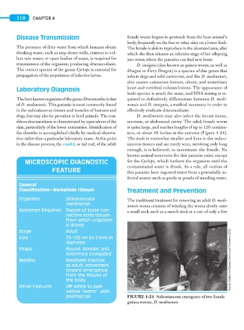
skin, particularly of the lower extremities. Identification of is quite large, and reaches lengths of up to 120 centime-
the disorder is accomplished chiefly by medical observa- ters, or about 48 inches at the extreme (Figure 4-24).
tion rather than a particular laboratory exam. At this point The male is somewhat smaller and lives in the subcu-
in the disease process, the caudal, or tail end, of the adult taneous tissues and are rarely seen, surviving only long
enough, it is believed, to inseminate the female. No
known animal reservoirs for this parasite exist, except
MICROSCOPIC DIAGNOSTIC for the Cyclops, which harbors the organism until the
contaminated water is drunk. As a rule, all victims of
FEATURE this parasite have ingested water from a potentially in-
fected source such as pools or ponds of standing water.
General
Classification—Nematode (tissue) Treatment and Prevention
Organism Drancunculus The traditional treatment for removing an adult D. medi-
medinensis
nensis worm consists of winding the worm slowly onto
Specimen Required Papule of loose con- a small stick such as a match stick at a rate of only a few
nective body tissues
from which organism
is drawn
Stage Adult
Size 70–120 cm by 2 mm in
diameter
Shape Round, slender, and
extremely elongated
Motility Relatively inactive Source: Centers for Disease Control and Prevention (CDC)
as adult; movement
toward emergence
from the tissues of
the body
Other Features Off-white to pale
yellow “worm” with
pointed tail FIGURE 4-24 Subcutaneous emergence of two female
guinea worms, D. medinensis