
Page 81 - parasitology for medical and clinical laboratoryprofessionals
P. 81
Protozoal Microorganisms as Intestinal Parasites 61
MICROSCOPIC DIAGNOSTIC
FEATURE
General
Classification Amoeba Source: Centers for Disease Control and Prevention (CDC)
Organism Entamoeba
hartmanni
Specimen Required Stool specimen
Stage Trophozoites most
diagnostic stage
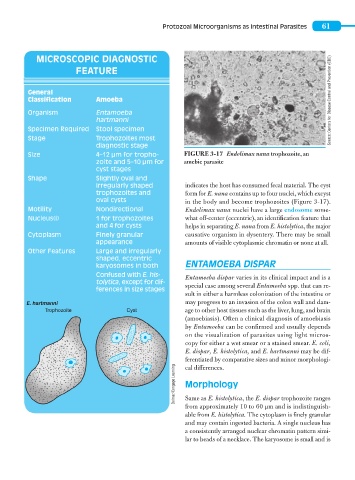
Size 4–12 μm for tropho- FIGURE 3-17 Endolimax nana trophozoite, an
zoite and 5–10 μm for amebic parasite
cyst stages
Shape Slightly oval and
irregularly shaped indicates the host has consumed fecal material. The cyst
trophozoites and form for E. nana contains up to four nuclei, which excyst
oval cysts in the body and become trophozoites (Figure 3-17).
Motility Nondirectional Endolimax nana nuclei have a large endosome some-
Nucleus(i) 1 for trophozoites what off-center (eccentric), an identification feature that
and 4 for cysts helps in separating E. nana from E. histolytica, the major
Cytoplasm Finely granular causative organism in dysentery. There may be small
appearance amounts of visible cytoplasmic chromatin or none at all.
Other Features Large and irregularly
shaped, eccentric
karyosomes in both ENTAMOEBA DISPAR
Confused with E. his- Entamoeba dispar varies in its clinical impact and is a
tolytica, except for dif-
ferences in size stages special case among several Entamoeba spp. that can re-
sult in either a harmless colonization of the intestine or
E. hartmanni may progress to an invasion of the colon wall and dam-
Trophozoite Cyst age to other host tissues such as the liver, lung, and brain
(amoebiasis). Often a clinical diagnosis of amoebiasis
by Entamoeba can be confirmed and usually depends
on the visualization of parasites using light micros-
copy for either a wet smear or a stained smear. E. coli,
E. dispar, E. histolytica, and E. hartmanni may be dif-
ferentiated by comparative sizes and minor morphologi-
Delmar/Cengage Learning Morphology
cal differences.
Same as E. histolytica, the E. dispar trophozoite ranges
from approximately 10 to 60 μm and is indistinguish-
able from E. histolytica. The cytoplasm is finely granular
and may contain ingested bacteria. A single nucleus has
a consistently arranged nuclear chromatin pattern simi-
lar to beads of a necklace. The karyosome is small and is