
Page 84 - parasitology for medical and clinical laboratoryprofessionals
P. 84
64 CHAPTER 3
species Entamoeba histolytica, the nonpathogenic species
Entamoeba dispar, and other species that occasionally
infect humans. Multiple samples often have to be re-
quested and examined as the presence of cysts of the
genera for Entamoeba, Iodamoeba, or Endolimax can
cause difficulty in making a diagnosis. In sporadic
cases of human infection with other species such as Source: Centers for Disease Control and Prevention (CDC)
E. moshkovskii accompanied with both E. histolytica and
E. dispar in young children in Bangladesh (Ali, et al.,
2003), the differentiation of the three species in clinical
samples by other means becomes of great importance
both for diagnosis and for epidemiological studies.
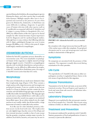
Although there is some evidence that following infection FIGURE 3-19 Iodamoeba buetschlii cyst, an amoebal
with E. dispar, pathological changes may occur in some parasite
humans (McMillan, et al., 1984), at this point E. dispar
is still largely considered a nonpathogen. the cytoplasm with a large karyosome that may fill much
of the nuclear space within the cytoplasm. No peripheral
chromatin may be seen and nonprogressive and slow
IODAMOEBA BUTSCHLII motility is characteristic of this species.
Iodamoeba butschlii organisms present themselves as Symptoms
mononuclear cysts. The most remarkable morphologi-
cal feature of this organism is a lightly stained and large No symptoms are associated with the presence of this
glycogen supply vacuole. I. butschlii is a nonpathogenic organism. The organism is usually discovered during
amoeba with worldwide distribution and prevalence. examinations for other parasites.
Although it can be difficult to differentiate the tropho-
zoites of I. butschlii from Endolimax nana, this stage of Life Cycle
I. butschlii is more active than that of E. nana.
The reproduction of I. butschlii is the same as other non-
pathogenic amoebae. Longitudinal binary fission is the
Morphology
sole means of reproduction for this organism.
The cysts of Iodamoeba are much more distinctive than
their trophozoite stage (Figure 3-19). They contain a Disease Transmission
single nucleus with a large karyosome and inconspicuous Transmission is by the fecal-oral route, as are most of the
peripheral chromatin. Cysts are variable in size but are
intestinal amoebae. Personal hygiene and ingestion of
mostly 5 to 20 μm in diameter and also contain the char- foods and water from safe sources will eliminate infec-
acteristic and well-defined large glycogen vacuole but no
tions by I. butschlii.
readily discernible chromatoidal bars. The large glycogen
vacuole stains a deep reddish-brown with iodine and in Laboratory Diagnosis
permanent stains the vacuole may appear as an unstained
intracellular space. In an iodine-stained cyst, the single Diagnosis is accomplished by the microscopic examina-
nucleus is not readily visible, but if discernible, a large tion of fecal samples for I. butschlii. Wet mounts using
karyosome is located eccentrically in the nucleus. solutions of iodine are effective in visualizing I. butschlii.
As is the case for other intestinal protozoa, infec-
tion occurs via the fecal-oral route. The trophozoites Treatment and Prevention
are only slightly larger than the cysts at 10 to 20 μm and
their internal structure is similar to that of cysts except No treatment is indicated for an infection with I. butschlii.
that the granular cytoplasm contains many vacuoles. In Sanitary preparation of food and purification of water is
addition, fecal debris, bacteria, and yeast may be seen in necessary to prevent the infection.