
Page 93 - parasitology for medical and clinical laboratoryprofessionals
P. 93
Protozoal Microorganisms as Intestinal Parasites 73
among those with normal immune systems. Microspo-
ridia have the potential to be waterborne because they
are released in both feces and urine that may wash into
bodies of water. Although the most frequent cause of
human microsporidium infection is Enterocytozoon
bieneusi. Other microsporidia that are well known
include: Encephalitozoon hellem, Encephalitozoon
cuniculi, Encephalitozoon intestinalis, and Nosema
corneum. Further information may be found on the fol-
lowing Internet site: http://www.dpd.cdc.gov/DPDx/
HTML/ImageLibrary/Microsporidiosis_il.htm.
Microsporidia may infect individuals through
both the digestive and the respiratory systems. Resis-
tant spores are formed within the host and then are
excreted from the body in feces and urine, and perhaps
by mucous secretion, but this route has not been fully Source: Centers for Disease Control and Prevention (CDC)
verified. Therefore, microsporidiosis is predisposed to
spread via fecal-oral, urine-oral, and waterborne trans-
mission. Microsporidia spores have been shown to
survive for protracted periods of time in water (up to
4 months) and have been detected in surface water
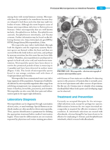
(Figure 3-22). The levels of microsporidia spores FIGURE 3-22 Microsporidia—electron micrograph of
found in raw sewage correlates well with those of a mature microsporidian spore
Cryptosporidium and Giardia.
Microsporidia from contaminated water may infect with Giemsa or Gram stains are not effective for detecting
large segments of the population. Symptoms of individu- spores in the presence of bacteria that is normally pres-
als infected by microsporidia organisms often range from ent in stool specimens and other sources of specimens.
exhibiting asymptomatic (no symptoms) conditions to But the use of a Giemsa stain is valuable when staining
bouts of diarrhea, bronchitis, pneumonia, and sinusitis. duodenal fluid where both spores and developing stages
Microsporidia can also cause bile duct pain and inflam- can be observed.
mation (pain in the upper-right abdomen).
Treatment and Prevention
Laboratory Diagnosis
Currently no accepted therapies for the microspo-
Microsporidiosis can be diagnosed through examination ridial infections exist, except for perhaps two species.
of stool, urine, or nasal washings. Special fluorescent an- Differentiation between the two major intestinal mi-
tibody studies are often needed to detect microsporidia. crosporidia is required for effective treatment of the
Thus, species identification is paramount for defining condition. E. intestinalis infections are treated with
the appropriate treatment before medical intervention albendazole, whereas fumagillin has been shown to be
begins. These tests for differentiation are not routinely effective for eradicating E. bieneusi, and Encephalitozoon
requested by physicians and routine staining procedures intestinalis, which is treated with albendazole.