
Page 22 - Tobillo y Pie 9.1
P. 22
Posterior ankle arthroscopic approach for the treatment of Raikin´s 7-8-9 osteochondral lesions of the talus
Clinical evaluation RESULTS
The results were evaluated using the AOFAS clinical The mean preoperative AOFAS score was 45.5
(10)
rating system for the ankle-hindfoot. Four questions points (33-70) and the mean postoperative score was
were also asked at the final evaluation: 85.29 (60-100). All patients satisfied with the outcome
1. Does the patient satisfy about the surgical outcome? of operation and would undergo the same operation.
2. Will the patient undergo the procedure again? The average VAS improved from preoperative 7.75
3. Is there pain at the portal scars? points to postoperative 1.54 points with an average
4. Has the patient returned to pre-injury physical improvement of 6.21 points. All the patients returned
to sport activities but only 75% of the patients restored
activity level? the pre-injury activity level (Table 1 and 2).
Complementarily visual analogue scale (VAS) was
used to evaluate the clinical behavior of patients. Complications
There were two postoperative complications: one
Surgical technique patient presented a large ecchymosis and another
The patient was in prone position and the operation presented a transient hypoesthesia of the heel because
was performed under spinal anesthesia. Posterior ankle of injury to the calcaneal branch of the tibial nerve.
arthroscopy was performed with the posteromedial and
posterolateral portals. The posterior intermalleolar Twelve patients (50%) had induration and pain in
(4)
ligament was identified and cut. This allowed approach the portals during the first postoperative two months.
to the posterior ankle joint through a trapezoidal This situation is resolved spontaneously. A total of 6
window. The window was bordered by the FHL patients were unable to regain the level of physical
medially, the transverse ligament proximally, the activity prior to the injury.
posterior talo-fibular ligament laterally and the posterior
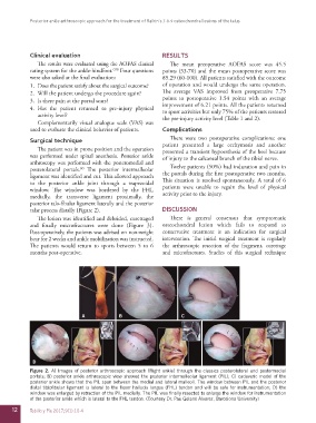
talar process distally (Figure 2). DISCUSSION
The lesion was identified and debrided, curettaged There is general consensus that symptomatic
and finally microfractures were done (Figure 3). osteochondral lesion which fails to respond to
Post-operatively, the patients was advised on non-weight conservative treatment is an indication for surgical
bear for 2 weeks and ankle mobilization was instructed. intervention. The initial surgical treatment is regularly
The patients would return to sports between 5 to 6 the arthroscopic resection of the fragment, curettage
months post-operative. and microfractures. Studies of this surgical technique
A B C
D
Figure 2. A) Images of posterior arthroscopic approach (Right ankle) through the classics posterolateral and postermedial
portals; B) posterior ankle arthroscopic view showed the posterior intermalleolar ligament (PIL); C) cadaveric model of the
posterior ankle shows that the PIL span between the medial and lateral malleoli. The window between PIL and the posterior
distal tibiofibular ligament is lateral to the flexor hallucis longus (FHL) tendon and will be safe for instrumentation; D) the
window was enlarged by retraction of the PIL medially. The PIL was finally resected to enlarge the window for instrumentation
of the posterior ankle which is lateral to the FHL tendon. (Courtesy Dr. Pau Golanó Alvarez, Barcelona University)
12 Tobillo y Pie 2017;9(1):10-4