Page 23 - Tobillo y Pie 9.1
P. 23

Batista JP, del Vecchio JJ, Maestu R, Patthauer L, Logioco LD, Lui TH


                                                                 Posterior ankle arthroscopy is a better approach
                                                              when treating these posterior lesions. This procedure,
                                                              in prone position, is a widely adopted approach that
                                                              can offer an excellent and secure arthroscopic view of
                                                              the posterior ankle. (1,4,9,14,15)
                                                                 Experience in posterior ankle arthroscopy and
                                                              knowledge of anatomy of the posterior ankle play a
                                                              fundamental role to a success treatment of the posterior
             A                       B
                                                              osteochondral lesions. Two ligaments can be identified
                                                              in the posterior ankle: the posterior tibiofibular ligament,
                                                              with its superficial and deep fascicles (Transverse ligament)
                                                              and the posterior talofibular ligament with its accessory
                                                              intermalleolar ligament.  The posterior intermalleolar
                                                                                  (11)
                                                              ligament is a ligament of the posterior tibiotalar joint that
                                                              is present in 56-100% of population. (16,17)  It is the source
                                                              of pain in some cases of posterior impingement syndrome.
                                                              The resection of this ligament creates a trapezoidal window
             C                       D                        for arthroscopic approach to the posterior ankle joint. This
                                                              provides adequate access for treatment of osteochondral
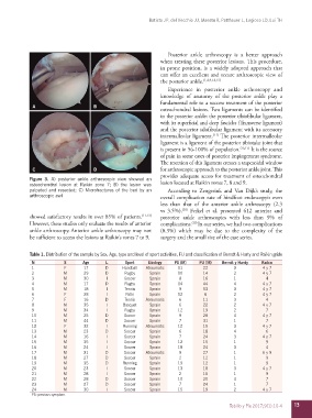
            Figure 3.  A)  posterior ankle  arthroscopic view  showed  an
            osteochondral lesion at Raikin zone 7; B) the lesion was   lesion located at Raikin zones 7, 8 and 9.
            palpated and resected; C) Microfractures of  the  bed by an   According to Zengerink and  Van Dijk’s study, the
            arthroscopic awl                                  overall complication rate of hindfoot endoscopyis even
                                                              less than that of the anterior ankle arthroscopy (2.3
                                                              vs 3.5%).  Ferkel et al. presented 612 anterior and
                                                                       (18)
            showed satisfactory results in over 85% of patients. (11-13)    posterior  ankle arthroscopies  with  less  than  9%  of
            However, these studies only evaluate the results of anterior   complications. (19)  In our series, we had two complications

            ankle arthroscopy. Anterior ankle arthroscopy may not   (8.3%) which may be due to the complexity of the
            be sufficient to access the lesions at Raikin’s zones 7 to 9.  surgery and the small size of the case series.


            Table 1. Distribution of the sample by Sex, Age, type and level of sport activities, FU and classification of Berndt & Harty and Raikin gride
             N           S       Age     L       Sport    Etiology  PS (M)   FU (M)   Berndt y Hardy  Raikin
             1            F      17      D      Handball  Atraumatic  11       22         3          4 y 7
             2           M       29      D       Rugby    Sprain     10        14         2          4 y 7
             3           M       30       I     Soccer    Sprain     8         16         1           4
             4           M       17      D       Rugby    Sprain     14        44         4          4 y 7
             5           M       18       I      Tennis   Sprain     9         53         3          4 y 7
             6            F      28       I      Patin    Sprain     12        6          2          4 y 7
             7            F      16      D       Tennis  Atraumatic  6         11         3           4
             8           M       35       I     Basquet   Sprain     6         22         2          4 y 7
             9           M       34       I      Rugby    Sprain     12        19         2           7
             10          M       25      D      Soccer    Sprain     9         28         4          4 y 7
             11          M       44      D      Soccer    Sprain     7         31         1           7
             12           F      32       I     Running  Atraumatic  12        15         3          4 y 7
             13          M       23      D      Soccer    Sprain     6         18         4           6
             14          M       16       I     Soccer    Sprain     7         24         3          4 y 7
             15          M       35       I     Soccer    Sprain     12        15         1           9
             16          M       24       I     Soccer    Sprain     18        24         3           4
             17          M       31      D      Soccer   Atraumatic  9         27         1          6 y 9
             18          M       27      D      Soccer    Sprain     2         12         1           9
             19          M       35      D      Running   Sprain     19        12         1           9
             20          M       23       I     Soccer    Sprain     13        18         3          4 y 7
             21          M       28       I     Soccer    Sprain     2         15         1           9
             22          M       28      D      Soccer    Sprain     10        20         4           7
             23          M       27      D      Soccer    Sprain     7         24         1           7
             24          M       30       I     Soccer    Sprain     15        19         2          4 y 7
             PS: previous symptom.

                                                                                         Tobillo y Pie 2017;9(1):10-4  13
   18   19   20   21   22   23   24   25   26   27   28