
Page 21 - Tobillo y Pie 9.1
P. 21
Batista JP, del Vecchio JJ, Maestu R, Patthauer L, Logioco LD, Lui TH
INTRODUCTION treatment for a minimum of six months. Fifteen
Osteochondral injuries of the ankle are relatively patients presented with locking symptoms of the ankle
rare lesions that primarily involve the cartilage and and none had sign of posterior ankle impingement. All
subchondral bone of the talus, and are presented with a patients had limitation in sporting activities or even
variable incidence ranging from 0.09 to 4%. It usually cannot participate the usual sports. Most had neither
(1)
presents with pain and disability during sports activities. local tenderness nor swelling, and none had a positive
Some patients experience pain during activities of daily anterior drawer test or a talar tilt test. The range of
living. Disputes still remain about the etiology and motion of the diseased ankle was comparable to the
(2)
pathogenesis of these lesions. (1-4) contralateral side. There was a reduction of sagittal
Ankle sprains and residual instability, are the most motion of less than 5º in 20 patients.
widely accepted etiology which has received various The treatment approach depends on the patient’s
terminology, e.g. osteochondral lesions, osteochondral symptoms (duration, rest pain and/or pain on exertion)
defects, trans chondral fractures, osteochondritis dissecans and the size and location of the defect. Whether they
and intraarticular fracture. (3,4) were large (about 15mm) and symptomatic and/or
There are classifications based on plain radiograph, unstable we opted for surgical treatment (2 patients who
CT and MRI. Based on these classifications, different remained in rehabilitation treatment for two months
prospects of the lesion can be assessed and accurate only). Intra-articular injections (e.g. corticosteriod,
surgical planning can be acchived. (4,5) Different hyaluronic acid, etc.) were not utilized prior to surgery.
treatment options have been proposed including We excluded patients with previous surgery to the
conservative and surgical treatment. Surgical treatment ankle, patients with Rheumatoid arthritis, joint
can be either arthroscopic and open surgery including (9)
debridement with or without microfractures, (6,7) impingement and/or ankle osteoarthritis.
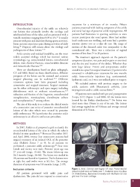
reduction and fixation of the fragment, osteochondral All patients were studied with pre and postoperative
transplantation, mosaicoplasty, chondrocyte culture X-rays, (CT) (Figure 1) and MRI. All the lesions were
(5)
(8)
and transplantation, among others. located by the Raikin classification. No lesion was
The aim of this study is to evaluate the clinical results sized more than 15mm in any of its axis. The lesions
of a consecutive case series of posterior osteochondral had average sagittal size of 9.16mm and average coronal
lesions of the talus which were treated by posterior dimension of 8.51mm.
ankle arthroscopy. We hypothesize that posterior ankle
arthroscopy is an effective and secure procedure.
METHODS
In 2007, Raikin et al. proposed a grid to classify the
osteochondral lesions of the talus in which the talus is
(5)
divided in nine quadrants. Between December 2011
and April 2004, twenty four consecutive patients had
surgical treatment of an isolated OCL of the posterior
talus (Raikin’s Zones 7 to 9). In all patients we A B
performed a posterior ankle arthroscopy. Four were
(4)
female and twenty were male. Patients’ mean age was
27 year-old (range, 16-44). The mean follow-up was
26.2 months (range, 18-84). The lesion was medial in
18 patients and lateral in 4 patients. Twelve lesions were
at right feet and twelve were at left feet. The average
duration of symptoms was 9.8 months (range, 2-19). C D
Inclusion criteria Figure 1. Axial, Coronal and Sagital CT view. Posteromedial
osteochondral lesion at Raikin zone 7. Anatomic piece (courtesy
All patients presented with ankle pain and Dr. Micki Dalmau, Barcelona University) with the Raikin & Elias
dysfunction. The pain did not respond to conservative grid over the talar cartilage
Tobillo y Pie 2017;9(1):10-4 11