
Page 24 - Tobillo y Pie 9.1
P. 24
Posterior ankle arthroscopic approach for the treatment of Raikin´s 7-8-9 osteochondral lesions of the talus
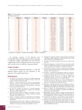
Table 2. The preoperative and postoperative AOFAS scores, VAS, size and type of treatment in patients treated through posterior
arthroscopic approach
N AOFAS pre AOFAS post VAS pre VAS post Size (mm) Uni or Bi Treat.
1 36 88 9 1 12 x 9,4 Unilateral DCM
2 43 80 8 2 8,5 x 11,2 Unilateral DCM
3 33 80 9 1 3,1 x 4,2 Unilateral DCM
4 55 90 9 1 8,4 x 13,1 Unilateral DC
5 44 60 7 3 13,2 x 10,5 Unilateral DC
6 40 78 8 3 9,2 x 11,1 Unilateral DCM
7 36 90 9 0 8,7 x 12,3 Bilateral DCM
8 40 86 8 0 7,23 x 10,6 Unilateral DC
9 48 88 8 0 10,3 x 8,3 Unilateral DC
10 40 90 8 1 12,2 x 9,6 Unilateral DC
11 36 74 9 1 7,8 x 6,4 Unilateral DCM
12 33 93 9 0 14,6 x 9,9 Bilateral DCM
13 58 100 6 0 8,4 X 9 ,1 Unilateral DCM
14 54 83 7 1 11 x 9 Unilateral DC
15 42 88 8 2 6,3 X 5,8 Unilateral DC
16 57 88 6 3 10,3 X 8,7 Unilateral DC
17 70 90 8 2 7,5 x 6 Unilateral DC
18 58 93 6 3 6,5 X 5,9 Unilateral DC
19 33 92 7 2 8,5 x 6,3 Unilateral DC
20 56 90 6 0 11,4 x 9,5 Unilateral DCM
21 44 74 8 2 7,4 X 5,2 Unilateral DC
22 40 86 9 3 8,7 x 6,4 Unilateral DC
23 63 88 6 3 8,6 X 7,3 Unilateral DCM
24 33 78 8 3 10,2 x 8,5 Unilateral DCM
7,75 1,54167
D: debridment; DC: debridment + curettage; DCM: debridment + curettage + microfractures.
In summary, resection of the posterior talar 8. Schneider TE, Karaikudi S. Matrix-Induced Autologous Chondrocyte
osteochondral lessions through posterior ankle arthroscopy Implantation (MACI) grafting for osteochondral lesions of the
talus. Foot Ankle Int. 2009;30(9):810-4.
is technically simple, reproducible and safe with few 9. Van Dijk CN. Hindfoot endoscopy. Foot Ankle Clin. 2006;11(2):
complications, thus reducing the time of return to sport 391-414.
and/or work activity in relation to open procedures. 10. Kitaoka HB, Alexander IJ, Adelaar RS, Nunley JA, Myerson MS,
Sanders M. Clinical rating systems for the ankle-hindfoot, midfoot,
hallux, and lesser toes. Foot Ankle Int. 1994;15(7):349-53.
CONCLUSIONS 11. Lee KB, Bai LB, Chung JY, Seon JK. Arthroscopic microfracture
The posterior arthroscopic approach is a safe and for osteochondral lesions of the talus. Knee Surg Sports Traumatol
effective surgical option for the treatment of the Arthrosc. 2010;18(2):247-53.
posterior talar osteochondral lesion. 12. Thermann H. Treatment of osteochondritis dissecans of the talus:
a long-term follow-up. Sports Med Arthrosc Rev. 1994;2:284-8.
REFERENCES 13. Zengerink M, Struijs PA, Tol JL, van Dijk CN. Treatment of
osteochondral lesions of the talus: a systematic review. Knee
1. van Dijk CN, de Leeuw PAJ, Scholten PE. Hindfoot endoscopy Surg Sports Traumatol Arthrosc. 2010;18(2):238-46.
for posterior ankle impingement. Surgical technique. J Bone Joint 14. Smyth NA, Zwiers R, Wiegerinck JI, Hannon CP, Murawski CD,
Surg Am. 2009;91 Suppl 2:287-98. van Dijk CN, Kennedy JG. Posterior hindfoot arthroscopy: a
2. Berndt A, Harty M. Transchondral fractures (osteochondritis review. Am J Sports Med. 2014;42(1):225-34.
dissecans) of the talus. J Bone Joint Surg Am. 1959;41:988-1020. 15. Yoshimura I, Naito M, Kanazawa K, Ida T, Muraoka K, Hagio T.
3. Stone JW. Osteochondral lesions of the talar dome. J Am Acad Assessing the safe direction of instruments during posterior ankle
Orthop Surg. 1996;4(2):63-73. arthroscopy using an MRI model. Foot Ankle Int. 2013;34(3):434-8.
4. van Dijk CN, Scholten PE, Krips R. A 2-portal endoscopic 16. Golanó P, Mariani PP, Rodríguez-Niedenfuhr M, Mariani PF,
approach for diagnosis and treatment of posterior ankle pathology. Ruano-Gil D. Arthroscopic anatomy of the posterior ankle ligaments.
Arthroscopy. 2000;16(8):871-6. Arthroscopy. 2002;18(4):353-8.
5. Bozkurt M, Yilmaz E, Atlihan D, Tekdemir I, Havitçioğlu H, Günal I. 17. Rosenberg ZS, Cheung YY, Beltran J, Sheskier S, Leong M, Jahss M.
The proximal tibiofibular joint: an anatomic study. Clin Orthop Posterior intermalleolar ligament of the ankle: normal anatomy and
Relat Res. 2003;(406):136-40. MR imaging features. AJR Am J Roentgenol. 1995;165(2):387-90.
6. Becher C, Thermann H. Results of microfracture in the treatment 18. Zengerink M, van Dijk CN. Complications in ankle arthroscopy.
of articular cartilage defects of the talus. Foot Ankle Int. 2005; Knee Surg Sports Traumatol Arthrosc. 2012;20(8):1420-31.
26(8):583-9. 19. Ferkel RD, Zanotti RM, Komenda GA, Sgaglione NA, Cheng MS,
7. Chuckpaiwong B, Berkson EM, Theodore GH. Microfracture for Applegate GR, Dopirak RM. Arthroscopic treatment of chronic
osteochondral lesions of the ankle: outcome analysis and outcome osteochondral lesions of the talus: long-term results. Am J Sports
predictors of 105 cases. Arthroscopy. 2008;24(1):106-12. Med. 2008;36(9):1750-62.
14 Tobillo y Pie 2017;9(1):10-4