Page 288 - 00. Complete Version - Progress Report IPEN 2014-2016
P. 288
288   Materials and Nanotechnology | Progress Report


               Biomaterials                                   active glass-ceramic powder was synthesized

                                                              by a modified sol-gel method. Non-aggregat-
               CaO-MgO-SiO2 ceramic glass system              ed SiO2 particles were initially prepared by
                                                              surfactant template sol-gel technique, via
                                                              acid-catalyzed hydrolysis from Na2SiO3. Ca
                                                              and Mg ions were embedded on the silica
                                                              particles. The glass-ceramic body sintered
                                                              at 1320 οC, from synthesized powder, con-
                                                              firmed the presence of bioactive phases, such
                                                              as, CaSiO3 and Ca2MgSi2O7. Immersion stud-
                                                              ies in simulated body fluid (SBF) for different
                                                              time intervals were carried out to study the
                                                              bioactivity of the material. The evolution of
                                                              bioactivity revealed, after 7 days soaking of
                                                              the glass-ceramic body in SBF, apatite parti-
                                                              cles were formed on the surface, confirmed by
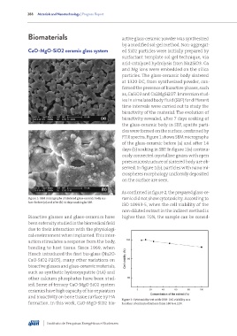
                                                              FTIR spectra. Figure 1 shows SEM micrographs
                                                              of the glass-ceramic before (a) and after 14
                                                              days (b) soaking in SBF. In figure 1(a) continu-
                                                              ously connected crystalline grains with open
                                                              pores microstructure of sintered body are ob-
                                                              served. In figure 1(b), particles with nano mi-
                                                              crospheres morphology uniformly deposited
                                                              on the surface are seen.


                                                              As confirmed in figure 2, the prepared glass-ce-
               Figure 1: SEM micrographs of sintered glass-ceramic body sur-  ramic did not show cytotoxicity. According to
               face: before (a) and after (b) 14 days soaking in SBF.
                                                              ISO 10993-5, when the cell viability of the
                                                              non-diluted extract in the indirect method is
               Bioactive glasses and glass-ceramics have      higher than 70%, the sample can be consid-
               been extensity studied in the biomedical field
               due to their interaction with the physiologi-
               cal environment when implanted. This inter-
               action stimulates a response from the body,
               bonding to host tissue. Since 1969, when
               Hench introduced the first bio-glass (Na2O-
               CaO-SiO2-P2O5), many other variations on
               bioactive glasses and glass-ceramic materials,
               such as synthetic hydroxyapatite (HA) and
               other calcium phosphates have been stud-
               ied. Some of ternary CaO-MgO-SiO2 system
               ceramics have high capacity of bio-reparation
               and bioactivity on bone tissue surface by HA
                                                              Figure 2: Cytotoxicity test with CHO: Cell viability as a
               formation. In this work, CaO-MgO-SiO2 bio-     function of extracts dilutions from 100 to 6.25%.


                         Instituto de Pesquisas Energéticas e Nucleares

