Page 291 - 00. Complete Version - Progress Report IPEN 2014-2016
P. 291
Materials and Nanotechnology | Progress Report  291


               tal hip prostheses.                            for the marking did not induce a decrease in
                                                              the cellular viability of the samples, as no cy-
               Laser bean marking applied                     totoxicity was showed even after prolonged
               to orthopedic implant                          incubation. The studied biomaterial was ad-
                                                              equate on the biomechanical tests, since the
                                                              laser treatments, under the conditions used,
                                                              did not induce the formation of surface ten-
                                                              sions of magnitude capable of leading the fa-
                                                              tigue fracture, indicating infinite fatigue life;
                                                              the region of fracture by tension could not be
                                                              related to the laser marking (Figure 6c). The
                                                              wear volume decreased as a function of the
                                                              increase in micro hardness produced by the
                                                              increase of the pulse frequency in the textur-
                                                              ing. The visual character of the markings and
                                                              texturing was assured after the majority of
                                                              the tests performed.


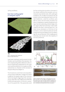
               Figure 5: Topography of (a) untreated and
               (b) textured laser biomaterials

               Laser beam marking is used to ensure the iden-
               tification and traceability of biomaterials. The
               texturing imparts greater adhesion to the sur-
               faces of implantable medical devices. The treat-
               ments performed altered the roughness of the
               biomaterials as a function of the increase of
               the pulse frequency (Figure 5)


               The microstructure and chemical composition
               of the surfaces underwent changes that direct-
               ly affected the passive layer of the stainless
               steels, favoring the triggering of the corrosion
                                                              Figure 6: (a) Regions of the sample-4 analyzed by points and by
               process. This effect was evidenced by SVET, XPS   line, respectively. The arrow indicates the direction adopted for
               and the characterization of electronic proper-  the analysis by “line scan”- No attack, (b) On-line mapping of the
                                                              distribution of the chemical elements present on the sample sur-
               ties of the passive film. The parameters used   face-4, (c) Marked and standard fractured tensile specimens.

