Page 290 - 00. Complete Version - Progress Report IPEN 2014-2016
P. 290
290   Materials and Nanotechnology | Progress Report


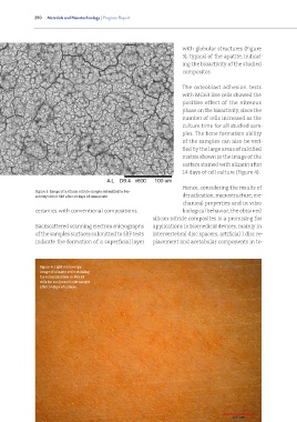
                                                                          with globular structures (Figure
                                                                          3), typical of the apatite, indicat-
                                                                          ing the bioactivity of the studied
                                                                          composites.


                                                                          The  osteoblast  adhesion  tests
                                                                          with MG63 line cells showed the
                                                                          positive  effect  of  the  vitreous
                                                                          phase on the bioactivity, since the
                                                                          number of cells increased as the
                                                                          culture time for all studied sam-
                                                                          ples. The bone formation ability
                                                                          of the samples can also be veri-
                                                                          fied by the large areas of calcified
                                                                          matrix shown in the image of the
                                                                          surface stained with alizarin after
                                                                          14 days of cell culture (Figure 4).


                                                                          Hence, considering the results of
               Figure 3: Image of a silicon nitride sample submitted to bio-
               activity test in SBF after 16 days of immersion            densification, microstructure, me-
                                                                          chanical properties and in vitro
               ceramics with conventional compositions.                   biological behavior, the obtained
                                                              silicon nitride composites is a promising for
               Backscattered scanning electron micrographs    applications in biomedical devices, mainly in
               of the samples surfaces submitted to SBF tests   intervertebral disc spacers, artificial l disc re-
               indicate the formation of a superficial layer   placement and acetabular components in to-


                 Figure 4: Light microscopy
                 image of alizarin red S staining
                 for mineralization in MG-63
                 cells for a silicon nitride sample
                 after 14 days of culture.


                         Instituto de Pesquisas Energéticas e Nucleares

