Page 295 - 00. Complete Version - Progress Report IPEN 2014-2016
P. 295
Materials and Nanotechnology | Progress Report  295


               facturing of parts with complex shapes and dimensions close
               to those of the end products (near-net shape process). The pro-
               duction of porous parts by space-holder technique using or-
               ganics comprises several steps, which are: (1) mixing organ-
               ic compounds with metal powder; (2) removal of the organic
               binder by heat treatment, generating the porous green body;
               (3) controlled sintering, in order to obtain porous structures
               with desirable mechanical properties. In this study, we aimed
               to produce porous implants of commercially pure titanium
               (cp-Ti) and Ti-13Nb-13Zr alloy by space-holder and suspension
               technique, using albumin as binder and natural polymers (corn
               starch, rice starch, potato starch and gelatin). The structure
               and surface features of the produced parts were analyzed by
               X-ray diffraction microscopy techniques. Special attention is
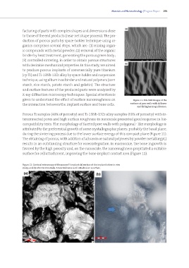
               given to understand the effect of surface nanoroughness on            Figure 11: FEG-SEM images of the
               the interaction between the implant surface and bone cells.         surfaces of pore walls with (a) lower
                                                                                       and (b) higher magnification.

               Porous Ti samples (40% of porosity) and Ti-13Nb-13Zr alloy samples (60% of porosity) with in-
               terconnected pores and high surface roughness in nanoscale presented good response in bio-
               compatibility tests. The morphology of faceted pore walls with polygonal - like morphology is
               attributed to the preferential growth of some crystallographic planes, probably the basal plane,
               during the sintering process due to the lower surface energy of this compact plane (Figure 11).
               The obtaining of porous, with addition of albumin or natural polymers by powder metallurgy(,)
               results in an outstanding structure for osseointegration. In macroscale, the bone ingrowth is
               favored by the high porosity and, on the nanoscale, the nanoroughness propitiated a suitable
               surface for cell attachment, improving the bone implant contact area (Figure 12).


               Figure 12: Confocal microscopy of the porous Ti implant: (a) surface of the implant before in vivo
               study; and (b) after in vivo study. Arrow indicates a cell attached on a surface.

