Page 297 - 00. Complete Version - Progress Report IPEN 2014-2016
P. 297
Materials and Nanotechnology | Progress Report  297


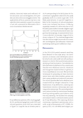
               solution, deionized water and methanol. Af-    26.3º corresponding to the (002) plane. On the
               ter sonication and centrifugation, rGO pow-    conversion to graphene oxide (GO), this peak
               ders are oven-dried and deagglomerated. The    gradually shifts to a lower angle (2ο= 9.1º).
               applicability of these powders has been eval-  Wide peak between 15 and 35º might be re-
               uated for electrocatalyst support for polymer-  lated to the formation of reduced graphene
               ic fuel cell, supercapacitor electrodes and zir-  oxide since methanol has shown a reducing
               conia bioceramics reinforcement.               capability towards GO. After reduction of GO,
                                                              the (002) plane shifted towards a higher angle
                                                              value (2ο= 24.5º) due to removal of some oxy-
                                                              gen functional groups. A representative trans-
                                                              mission electron microscopy image of rGO is
                                                              shown in figure 15. The observed fragmented
                                                              thin film feature might be a result of an exces-
                                                              sive centrifugation and ultrasonic treatment
                                                              used along washing and exfoliation steps.


                                                              Eletroceramics


                                                              In the 2014-2016 period, research work has
               Figure 14: Normalized X-ray diffraction patterns of   been devoted to two main areas: the devel-
               the starting graphite (G), synthesized graphene ox-  opment of ceramic solid electrolytes and in-
               ide (GO) and reduced graphene oxide (rGO).
                                                              termetallics for solid oxide fuel cells and basic
                                                              research on electric field assisted pressureless
                                                              sintering of electroceramics. The solid electro-
                                                              lytes that were studied were yttria, scandia and
                                                              scandia-ceria stabilized zirconia, strontium
                                                              and manganese-doped lanthanum gallate, yt-
                                                              trium-doped barium zirconate. The synthesis
                                                              techniques for preparing all these electroce-
                                                              ramics were solid state reaction, peroxo-oxi-
                                                              dant, spray pyrolysis and polymeric precursor.
                                                              The ceramic powders that were obtained were
                                                              characterized by nitrogen adsorption, X-ray
                                                              fluorescence, X-ray diffraction, scanning and
                                                              transmission electron microscopy and scan-
                                                              ning probe microscopy. The pressed powders
                                                              were sintered either by conventional heating
                                                              dwelling-cooling profiles, spark plasma, or by
               Figure 15: Transmission electron microscopy
               (TEM) images of reduced graphene oxide (rGO)   two-step sintering. The facilities for carrying
                                                              out research work on sintered ceramic pieces
               X-ray diffraction patterns of the starting graph-  are X-ray diffraction, FEG scanning electron
               ite (G), synthesized graphene oxide (GO) and   microscopy, impedance spectroscopy over a
               reduced graphene oxide (rGO) are presented     wide range of temperature (RT-1500K), fre-
               in figure 14. Graphite has an intense peak at   quency (0.01 Hz-140 MHz) and oxygen par-

