
Page 353 - Medicine and Surgery
P. 353
P1: FAW
BLUK007-07 BLUK007-Kendall May 25, 2005 18:18 Char Count= 0
Chapter 7: Tumours of the nervous system 349
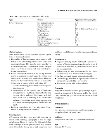
Table 7.16 Primary intracranial tumours and their frequency
Type Tumour Approximate frequency (%)
Primary malignancy Low-grade astrocytoma 5–8
Malignant astrocytoma 40
Oligodendroglioma 5
Ependymoma 10% in childhood, 1% in adults
Medulloblastoma 4
CNS lymphoma 3–5
Total (in all ages) ∼55
Benign Meningioma 20
Schwannoma (acoustic neuroma) 5–10
Pituitary tumour 10–20
Other, e.g. craniopharyngioma 7
Total (in all ages) ∼45
Clinical features and lower morbidity and mortality than standard open
Mass lesions within the skull produce signs and symp- biopsy.
toms by three mechanisms:
Direct effect of the mass causing compression or infil- Management
tration of the surrounding nervous tissue causes focal Surgical debulking may be performed. Complete re-
neurological signs. This may also occur secondary to section of benign tumours is preferred; however, if
surrounding oedema or arterial or venous compro- close to vital structures, e.g. brainstem lesions, this is
mise, i.e. a stroke. Headache with focal neurology is a not always possible.
typical presentation. Radiotherapy is used post-operatively, for unre-
Raised intracranial pressure (ICP) usually develops sectable lesions or in patients unfit for surgery.
slowly, so does not normally cause the typical triad Cerebral oedema is treated with corticosteroids.
of headache, vomiting and papilloedema. However, Chemotherapy is used for malignant astrocytoma, to
brainstem, floor of the third ventricle and cerebellar trytoprolong survival by a few months.
lesions tend to present initially with raised ICP: Seizures are treated with anticonvulsants.
i Distortion of the upper brain stem causes impaired
consciousness. Prognosis
ii Compression of the medulla due to herniation Prognosiscorrelateswithhistologictypeandgrade,post-
(coning) causes a third nerve lesion (due to com- operative size, extent of the tumour and by the patient
pression of the ipsilateral third nerve) and sixth characteristics (age, performance status, and duration of
nerve lesion (due to stretching of the contralateral symptoms).
sixth nerve), ipsilateral hemiparesis, impaired con-
sciousness, respiratory depression, bradycardia and Meningioma
death.
Partial or generalised tonic clonic seizures are charac- Definition
teristic of many cerebral mass lesions. Slow growing tumour arising from the meningeal cov-
ering of the brain and spinal cord.
Investigations
CT scanning will detect over 95% of intracranial tu- Incidence
mours. MRI scanning, angiography is used for surgi- They account for ∼20% of all intracranial tumours.
cal planning. Biopsy is required for histological diag-
nosis, although a radiological diagnosis may be suffi- Age
cient. Stereotactic biopsy has a high diagnostic yield Peak age 40–70 years.