Page 354 - Medicine and Surgery
P. 354
P1: FAW
         BLUK007-07  BLUK007-Kendall  May 25, 2005  18:18  Char Count= 0


                   350 Chapter 7: Nervous system


                   Sex                                          Gliomas (astrocytomas and
                   F > M (2.5:1)                                oligodendrogliomas)
                                                                Definition
                   Pathophysiology                              Tumours with histological appearances of glial cells – the
                   Meningiomas may grow to a large size over a period of  scaffolding cells of the CNS.
                   years. Most are benign, with 10% behaving in a malig-     Astrocytomas have predominantly astrocytic cells.
                   nant fashion. If they arise close to the skull they may  Theyarecategorisedaccordingtotheirhistologicalap-
                   erode the bone. Meningiomas often occur along venous  pearance into low grade astrocytoma (WHO grade 2),
                   sinuses.                                       anaplastic astrocytoma (WHO grade 3) and glioblas-
                                                                  toma multiforme (WHO grade 4).
                                                                    Oligodendromas arise from the oligodendrocytes
                   Clinical features
                   Most are asymptomatic and detected incidentally on  (CNS myelinating cells) and usually behave as low-
                   neuroimaging. The most common presentation is a  grade tumours.
                                                                    Mixedglial tumours with astrocytic and oligoden-
                   seizure or slowly progressive focal neurology. Visual or
                                                                  droglial components occur and are termed oligoas-
                   hearing abnormalities may be present, depending on
                                                                  trocytoma.
                   the site. Frontal tumours cause changes in personality.
                   A parasagittal (falx) meningioma causes a characteris-
                                                                Aetiology
                   tic pattern of bilateral leg weakness mimicking a spinal
                                                                Astrocytomas occur anywhere in the brain. Oligoden-
                   cord lesion. Spinal meningiomas cause limb weakness
                                                                drogliomas tend to arise in the cerebral hemispheres.
                   and numbness.
                                                                Glioblastomas are the most aggressive pleomorphic type
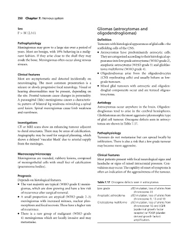
                                                                of glial cell tumour. Oncogene defects seen in astrocy-
                   Investigations                               tomas are shown in Table 7.17.
                   CT or MRI scans show an enhancing tumour adjacent
                   to dural structures. There may be areas of calcification.
                                                                Pathophysiology
                   Angiography may be used for surgical planning, which
                                                                Tumours do not metastasise but can spread locally by
                   shows a delayed ‘vascular blush’ due to arterial supply
                                                                infiltration. There is also a risk that a low grade tumour
                   from the meninges.
                                                                may become more aggressive.
                   Macroscopy/microscopy                        Clinical features
                   Meningiomas are rounded, rubbery lesions, composed  Most patients present with focal neurological signs and
                   of meningothelial cells with small foci of calcification  headache or signs of raised intracranial pressure. Con-
                   (psammoma bodies).                           vulsions may occur. The rapidity of onset of symptoms is
                                                                often an indication of the aggressiveness of the tumour.
                   Prognosis
                   Depends on histological features.
                                                                Table 7.17 Oncogene defects seen in astrocytomas
                     The vast majority are typical (WHO grade 1) menin-

                     giomas, which are slow growing and have a low risk  Low grade  p53 mutation, loss of alleles from
                     of recurrence after surgical removal.                           chromosome 22
                     A small proportion are atypical (WHO grade 2–3)
                                                                Anaplastic astrocytoma  p53 mutation, loss of alleles from
                                                                                     chromosome 9, 13 and 19
                     meningiomas with increased mitoses, nuclear pleo-  Glioblastoma multiforme  p53 mutation, loss of alleles from
                     morphism and focal necrosis. These have a higher rate           chromosome 10 and EGFR
                     of recurrence.                                                  (epidermal growth factor
                     There is a rare group of malignant (WHO grade
                                                                                     receptor) or PDGF (platelet
                                                                                     derived growth factor)
                     4) meningiomas which are locally invasive and may
                                                                                     amplification.
                     metastasise.

